Copyright
©2010 Baishideng.
World J Gastroenterol. Jun 14, 2010; 16(22): 2818-2823
Published online Jun 14, 2010. doi: 10.3748/wjg.v16.i22.2818
Published online Jun 14, 2010. doi: 10.3748/wjg.v16.i22.2818
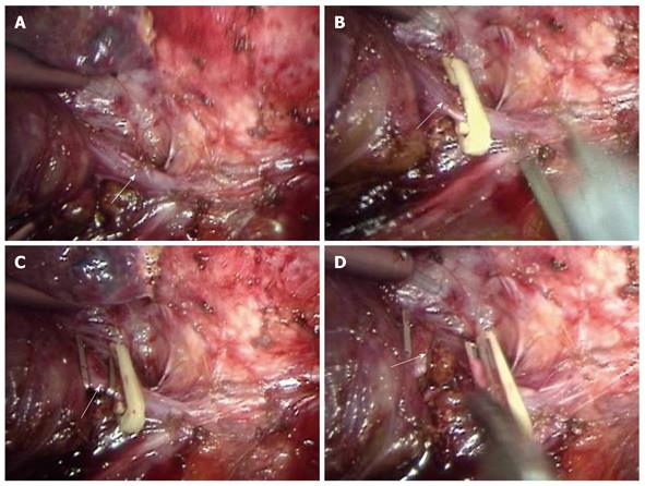
Figure 1 Laparoscopy showing dissected LHV (A, arrow), clamped LHV (B and C, arrow), and transected LHV (D, arrow).
LHV: Left hepatic vein.
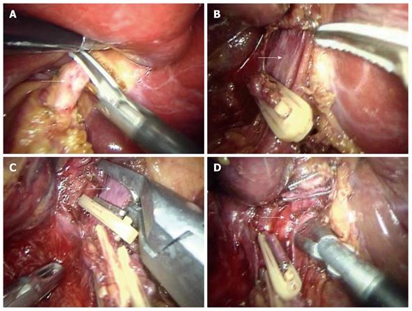
Figure 2 Laparoscopy showing dissected LBHA (A, arrow), exposed LBHV (B, arrow), dissected and clamped LBHV (C, arrow), and dissected left hepatic duct (D, arrow).
LBHA: Left branch of hepatic artery; LBHV: Left branch of hepatic vein.
-
Citation: Tu JF, Jiang FZ, Zhu HL, Hu RY, Zhang WJ, Zhou ZX. Laparoscopic
vs open left hepatectomy for hepatolithiasis. World J Gastroenterol 2010; 16(22): 2818-2823 - URL: https://www.wjgnet.com/1007-9327/full/v16/i22/2818.htm
- DOI: https://dx.doi.org/10.3748/wjg.v16.i22.2818


