©2010 Baishideng.
World J Gastroenterol. May 7, 2010; 16(17): 2120-2128
Published online May 7, 2010. doi: 10.3748/wjg.v16.i17.2120
Published online May 7, 2010. doi: 10.3748/wjg.v16.i17.2120
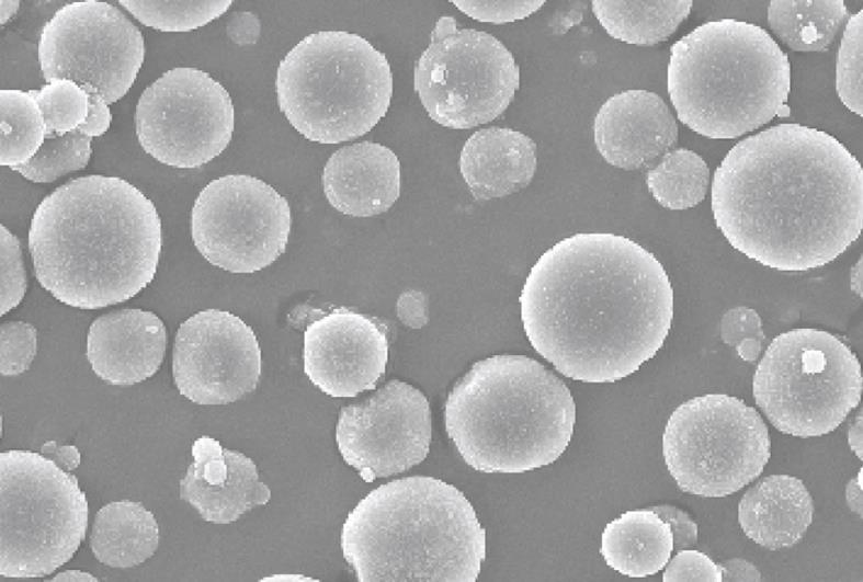
Figure 1 Scanning electron microscopic (20 kV) images of metal-coated gelatin microspheres (original magnification, × 500).
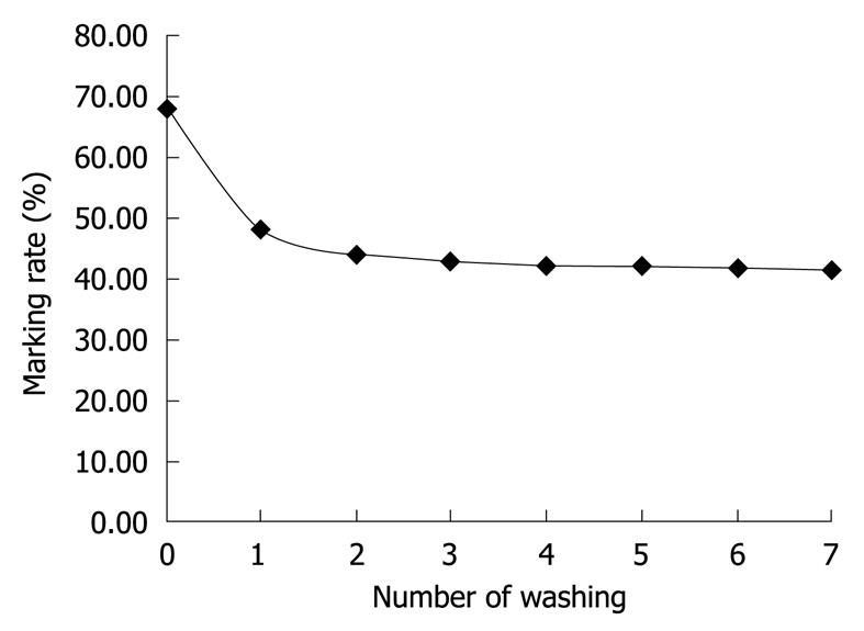
Figure 2 The labeling rate decreases with increasing the number of washes.
The slope decreased gradually, nearly reaching a straight line, which demonstrates that the 131I-labeled gelatin microspheres after washing contained very low amounts of nuclides conjugated by physical adsorption.
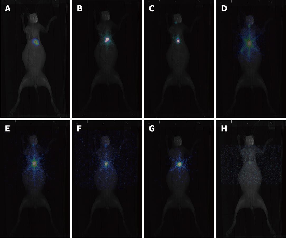
Figure 3 Single-photon emission computed tomography (SPECT) imaging performed at 4 h (A) and 1 (B), 4 (C), 8 (D), 16 (E), 24 (F), 32 (G) and 48 (H) d after the administration of 131I-labeled gelatin microspheres.
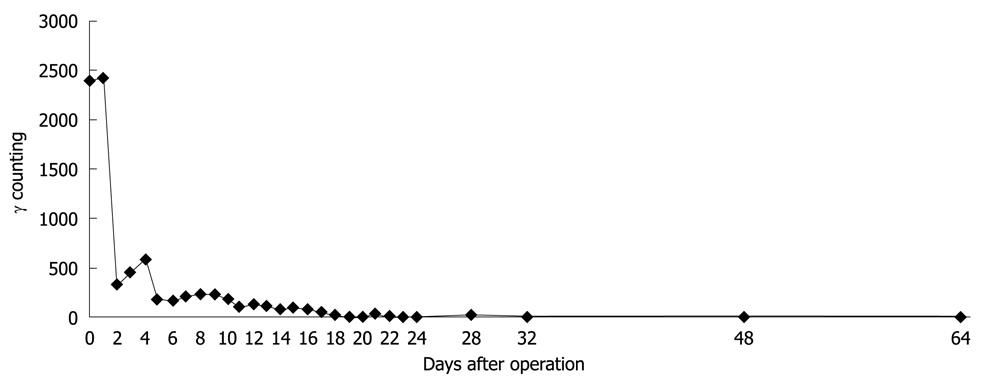
Figure 4 Dynamic changes in serum γ count after the administration of 131I-labeled gelatin microspheres.
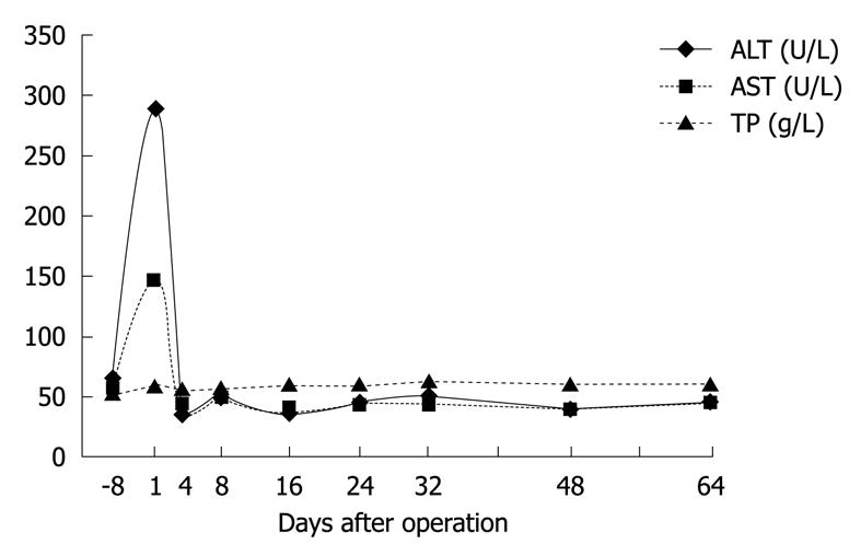
Figure 5 Changes in hepatic function after the administration of 131I-labeled gelatin microspheres.
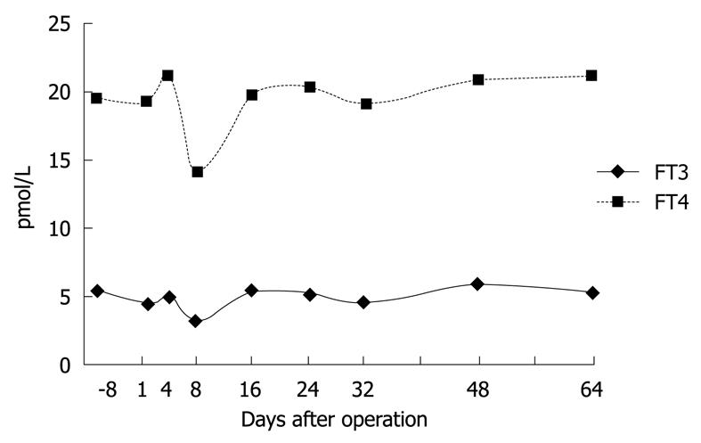
Figure 6 Curves of thyroid functional changes after the administration of 131I-labeled gelatin microspheres.
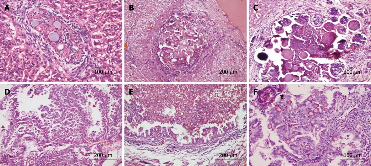
Figure 7 Pathological examination at 1 (A), 4 (B), 16 (C), 24 (D), 32 (E) and 48 (F) d after the administration of 131I-labeled gelatin microspheres (HE stain; original magnification, × 200 in B, C, D and E, and × 400 in A and F).
-
Citation: Ma Y, Wan Y, Luo DH, Duan LG, Li L, Xia CQ, Chen XL. Direct
in vivo injection of 131I-GMS and its distribution and excretion in rabbit. World J Gastroenterol 2010; 16(17): 2120-2128 - URL: https://www.wjgnet.com/1007-9327/full/v16/i17/2120.htm
- DOI: https://dx.doi.org/10.3748/wjg.v16.i17.2120


