©2010 Baishideng.
World J Gastroenterol. Mar 21, 2010; 16(11): 1422-1424
Published online Mar 21, 2010. doi: 10.3748/wjg.v16.i11.1422
Published online Mar 21, 2010. doi: 10.3748/wjg.v16.i11.1422
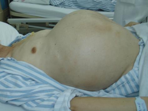
Figure 1 Patient had continuous abdominal uplift.
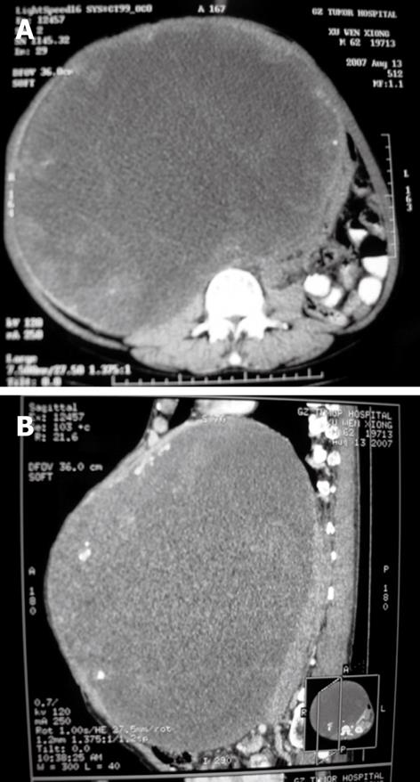
Figure 2 Computed tomography of the tumor.
A: A huge hepatic carcinoma at the right lobe of the liver. The left lobe of the liver was compensatory hypertrophy; B: A huge hepatic carcinoma above the right lobe of the liver and compression of the surrounding organs.
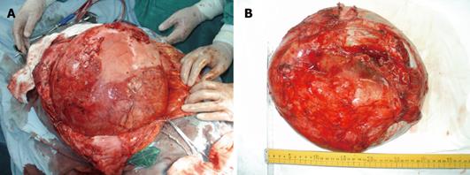
Figure 3 Imaging of the huge tumor.
A: The tumor was encompassed by a pseudocapsule and compressed surrounding organs; B: Postoperative measurement of tumor was 35 cm × 30 cm × 15 cm in size and 10 050 g in weight.
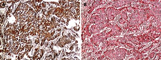
Figure 4 Pathological and biochemical examinations of the huge tumor.
A: Pathological examination showed the tumor was a well-differentiated HCC. HE × 400; B: Immunohistochemical staining showed the tumor was α-fetoprotein positive (++). HE × 400.
- Citation: Ba MC, Cui SZ, Lin SQ, Tang YQ, Wu YB, Zhang XL. Resection of a giant hepatocellular carcinoma weighing over ten kilograms. World J Gastroenterol 2010; 16(11): 1422-1424
- URL: https://www.wjgnet.com/1007-9327/full/v16/i11/1422.htm
- DOI: https://dx.doi.org/10.3748/wjg.v16.i11.1422


