©2010 Baishideng.
World J Gastroenterol. Jan 7, 2010; 16(1): 119-122
Published online Jan 7, 2010. doi: 10.3748/wjg.v16.i1.119
Published online Jan 7, 2010. doi: 10.3748/wjg.v16.i1.119
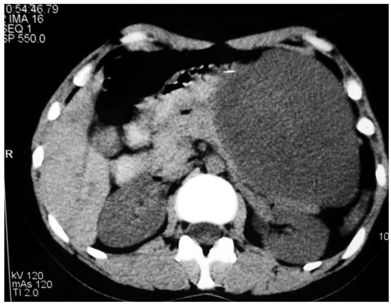
Figure 1 Computed tomography (CT) of pancreatic schwannoma with involvement of the transverse colon.
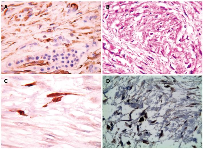
Figure 2 Microscopy of tumor cells infiltrating the serosa of the transverse colon.
A: Schwannoma pattern of growth: Antoni B type. HE, × 200; B: Characteristic elongated cells with cytoplasmic processes in area with loose, myxoid background. Immunohistochemical ABC method, × 400; C: Pancreatic schwannoma with subtotal pancreatic tissue. Immunohistochemical ABC method, × 400; D: Intense nuclear staining for Ki67 (MIB1) in the neoplastic cells. Immunohistochemical ABC method, × 400.
-
Citation: Stojanovic MP, Radojkovic M, Jeremic LM, Zlatic AV, Stanojevic GZ, Jovanovic MA, Kostov MS, Katic VP. Malignant schwannoma of the pancreas involving transversal colon treated with
en-bloc resection. World J Gastroenterol 2010; 16(1): 119-122 - URL: https://www.wjgnet.com/1007-9327/full/v16/i1/119.htm
- DOI: https://dx.doi.org/10.3748/wjg.v16.i1.119


