©2009 The WJG Press and Baishideng.
World J Gastroenterol. Mar 7, 2009; 15(9): 1138-1140
Published online Mar 7, 2009. doi: 10.3748/wjg.15.1138
Published online Mar 7, 2009. doi: 10.3748/wjg.15.1138
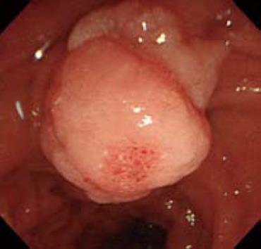
Figure 1 Endoscopy showing an 18-mm, whitish, elevated, slightly rough-surfaced mass, located proximal to the major papilla.
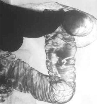
Figure 2 Hypotonic duodenography demonstrating a mass situated 15 mm proximal to the major papilla, which was raised highly from the duodenum.
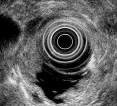
Figure 3 EUS detected an 18 mm × 12 mm homogeneous, hypoechoic mass in the submucosal layer.
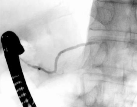
Figure 4 ERP was immediately performed via the minor papilla and it showed that the entire dorsal pancreatic ductal system was without communication with the ventral pancreatic duct.
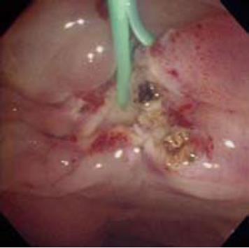
Figure 5 A pancreatic 5Fr stent was placed immediately after endoscopic papillectomy and coagulated the margin of the minor papilla tumor.
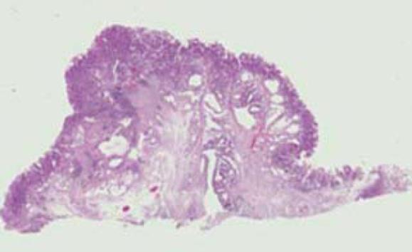
Figure 6 Histopathological findings of the specimen showed tubular adenoma and the margin of the tumor was negative; however, a slight infiltration of the pancreatic duct system was revealed (HE stain, × 4).
- Citation: Kanamori A, Kumada T, Kiriyama S, Sone Y, Tanikawa M, Hisanaga Y, Toyoda H, Kawashima H, Itoh A, Hirooka Y, Goto H. Endoscopic papillectomy of minor papillar adenoma associated with pancreas divisum. World J Gastroenterol 2009; 15(9): 1138-1140
- URL: https://www.wjgnet.com/1007-9327/full/v15/i9/1138.htm
- DOI: https://dx.doi.org/10.3748/wjg.15.1138


