Copyright
©2009 The WJG Press and Baishideng.
World J Gastroenterol. Dec 28, 2009; 15(48): 6123-6125
Published online Dec 28, 2009. doi: 10.3748/wjg.15.6123
Published online Dec 28, 2009. doi: 10.3748/wjg.15.6123
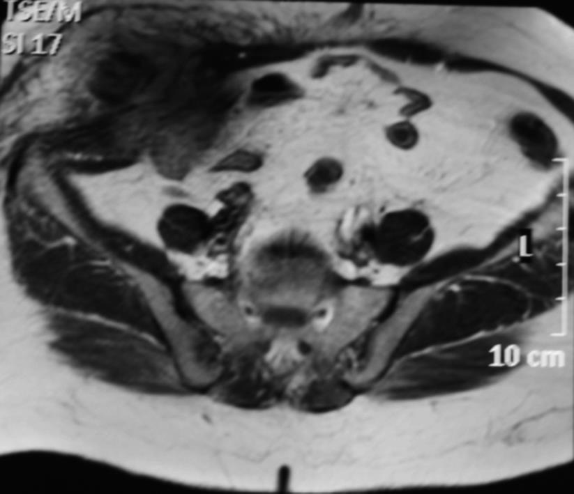
Figure 1 Pre-operative magnatic resonance imaging view of our case.
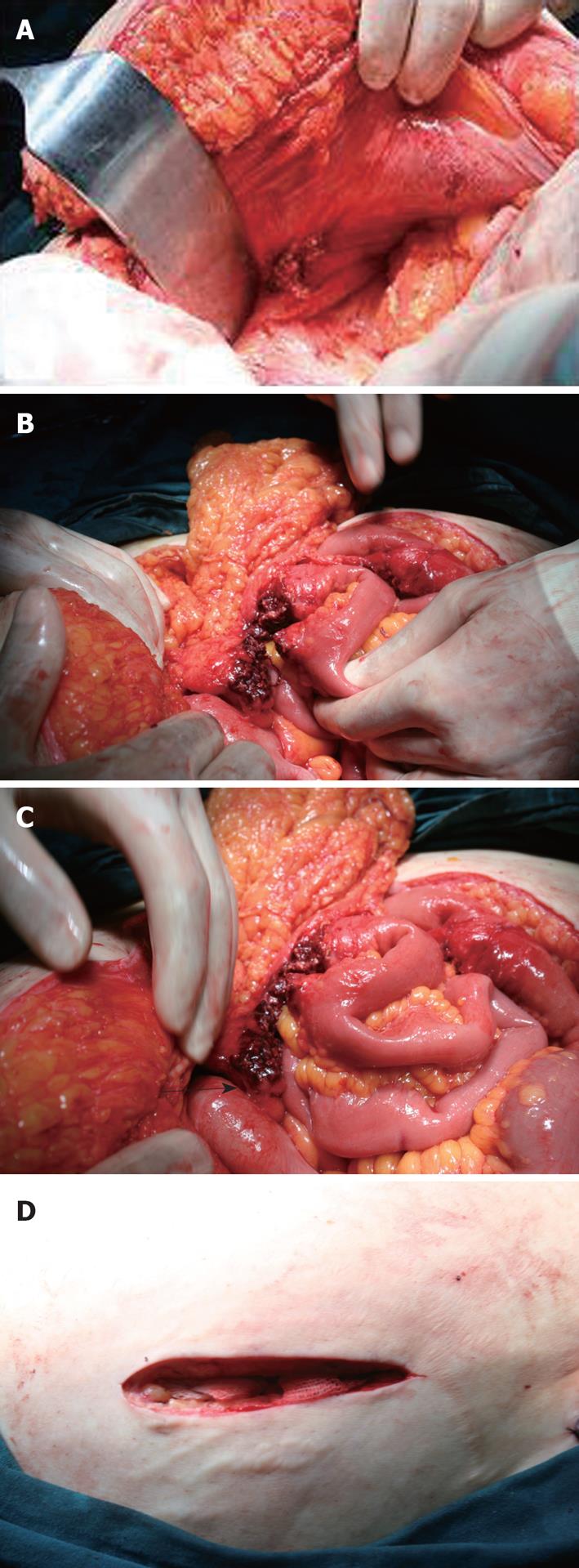
Figure 2 Intraoperative view of the meckel’s diverticulum manifested by cutaneous abscess.
A: The fistulization area of the diverticulum in the right lower abdominal wall; B: After retraction of the Meckel’s diverticulum: intraabdominal view of the diverticulum and necrotic omentum; C: The intraoperative view of the perforated diverticulum (black arrow shows Meckel’s diverticulum); D: The view of the abscess cavity.
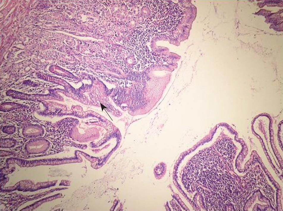
Figure 3 Meckel’s diverticulum (HE stain, × 100).
Photomicrograph shows the diverticulum composed of all layers of intestinal wall. Normal small intestinal mucosa and a focus of (arrow) gastric mucosa line the diverticulum.
- Citation: Karatepe O, Adas G, Altıok M, Ozcan D, Kamali S, Karahan S. Meckel’s diverticulum manifested by a subcutaneous abscess. World J Gastroenterol 2009; 15(48): 6123-6125
- URL: https://www.wjgnet.com/1007-9327/full/v15/i48/6123.htm
- DOI: https://dx.doi.org/10.3748/wjg.15.6123


