©2009 The WJG Press and Baishideng.
World J Gastroenterol. Nov 14, 2009; 15(42): 5368-5370
Published online Nov 14, 2009. doi: 10.3748/wjg.15.5368
Published online Nov 14, 2009. doi: 10.3748/wjg.15.5368
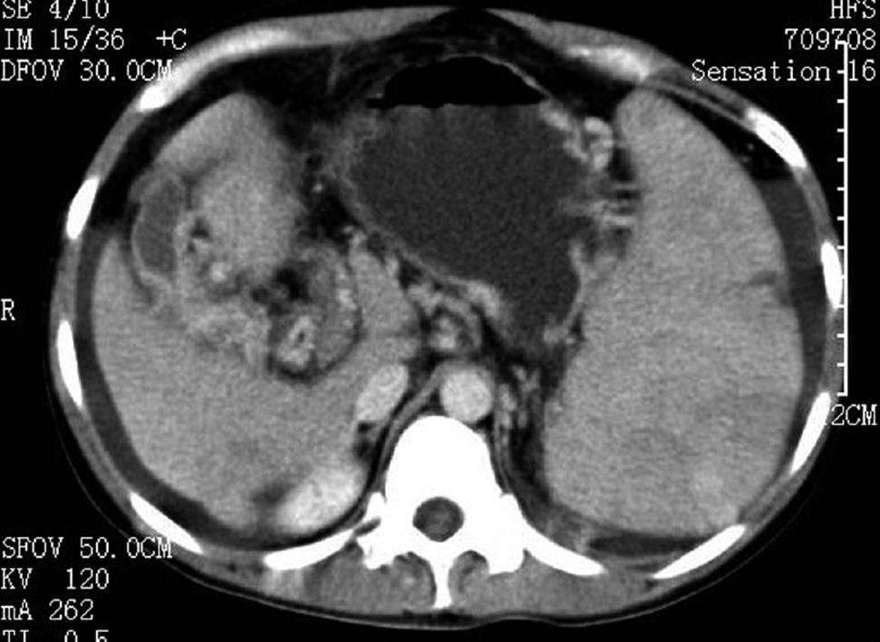
Figure 1 Computed tomography (CT) scan showed splenomegaly and varicose veins in the esophagus and gastric fundus.
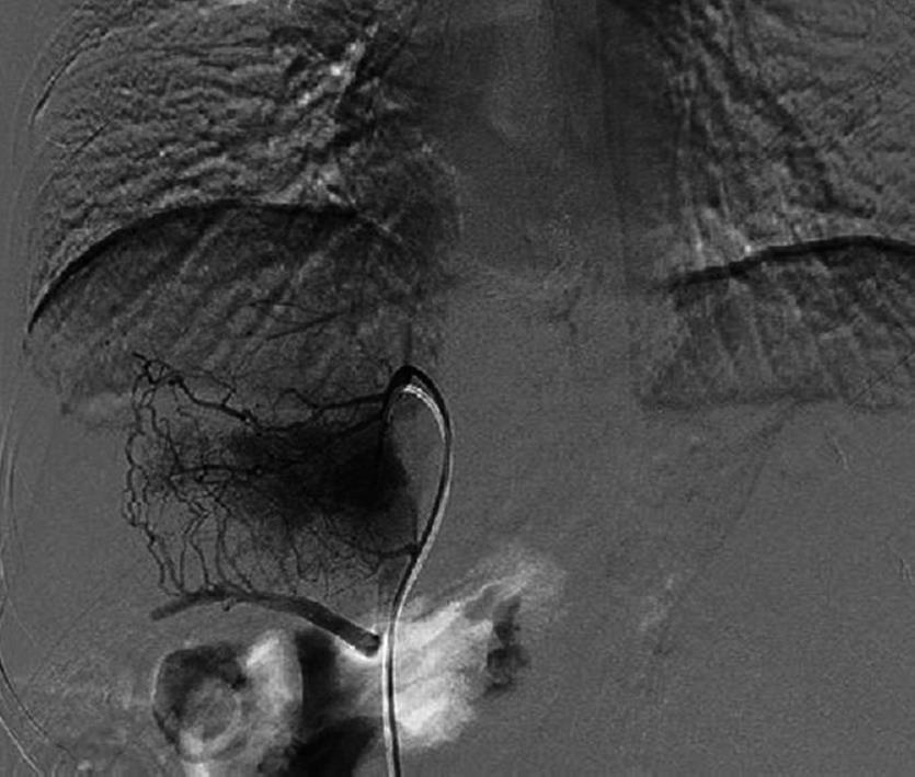
Figure 2 Portovenography showed non-visualization of the portal vein with formation of multiple collaterals in the hepatic hilum.
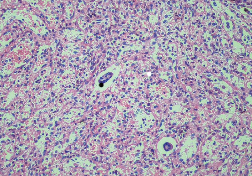
Figure 3 Spleen pathology showed chronic congestion and extramedullary hemopoiesis (magnification, × 200).
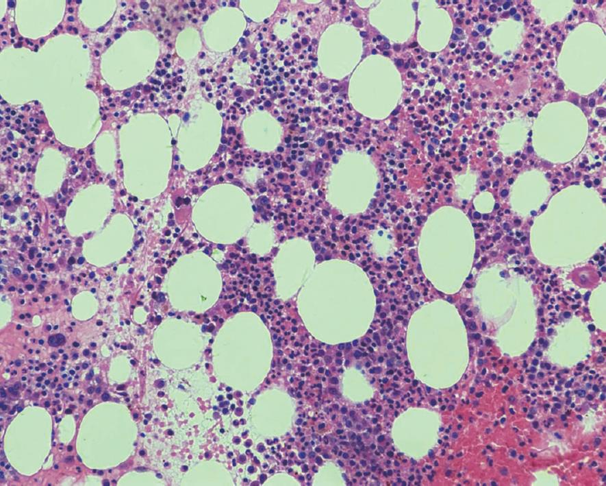
Figure 4 Bone marrow biopsy showed megakaryocytic hyperplasia (magnification, × 200).
- Citation: Cai XY, Zhou W, Hong DF, Cai XJ. A latent form of essential thrombocythemia presenting as portal cavernoma. World J Gastroenterol 2009; 15(42): 5368-5370
- URL: https://www.wjgnet.com/1007-9327/full/v15/i42/5368.htm
- DOI: https://dx.doi.org/10.3748/wjg.15.5368


