©2009 The WJG Press and Baishideng.
World J Gastroenterol. Nov 14, 2009; 15(42): 5307-5315
Published online Nov 14, 2009. doi: 10.3748/wjg.15.5307
Published online Nov 14, 2009. doi: 10.3748/wjg.15.5307
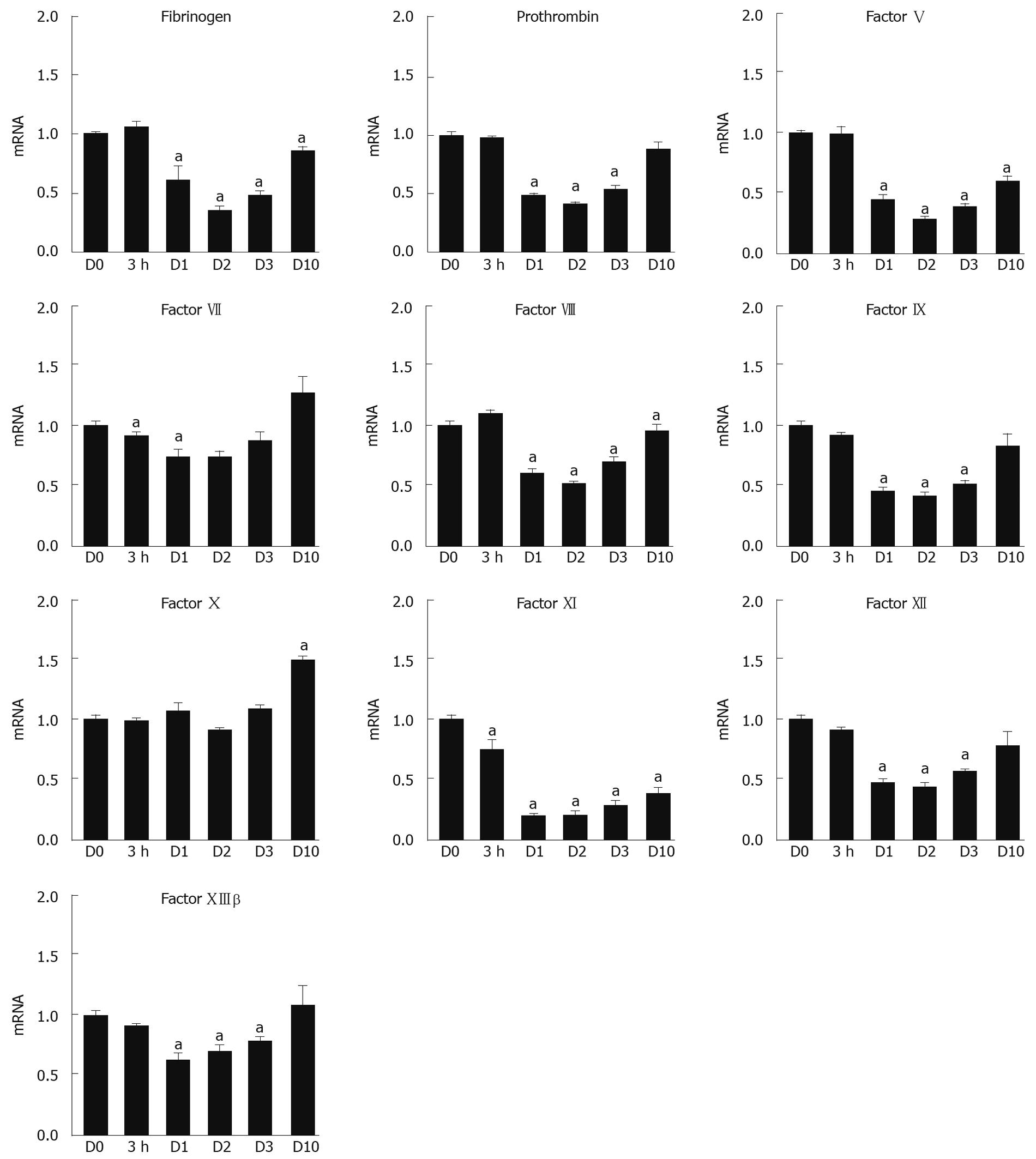
Figure 1 Gene expression profiling of coagulation factors in the liver during the DH-mediated regenerative phase following TCPOBOP induction.
The mRNA expression levels of 10 coagulation factors (fibrinogen, prothrombin, factors V, VII, VIII, IX, X, XI, XII, and XIIIβ) were assessed from mouse livers at D0, 3 h, D1, D2, D3, and D10 by real-time RT-PCR (n = 8/time point). The mRNA levels of each gene were normalized to the geometric mean of PPIA and RPL4 mRNA levels. The values were expressed as a comparative ratio to the day 0 samples, and represented as the mean ± SE. aP < 0.05 vs D0.
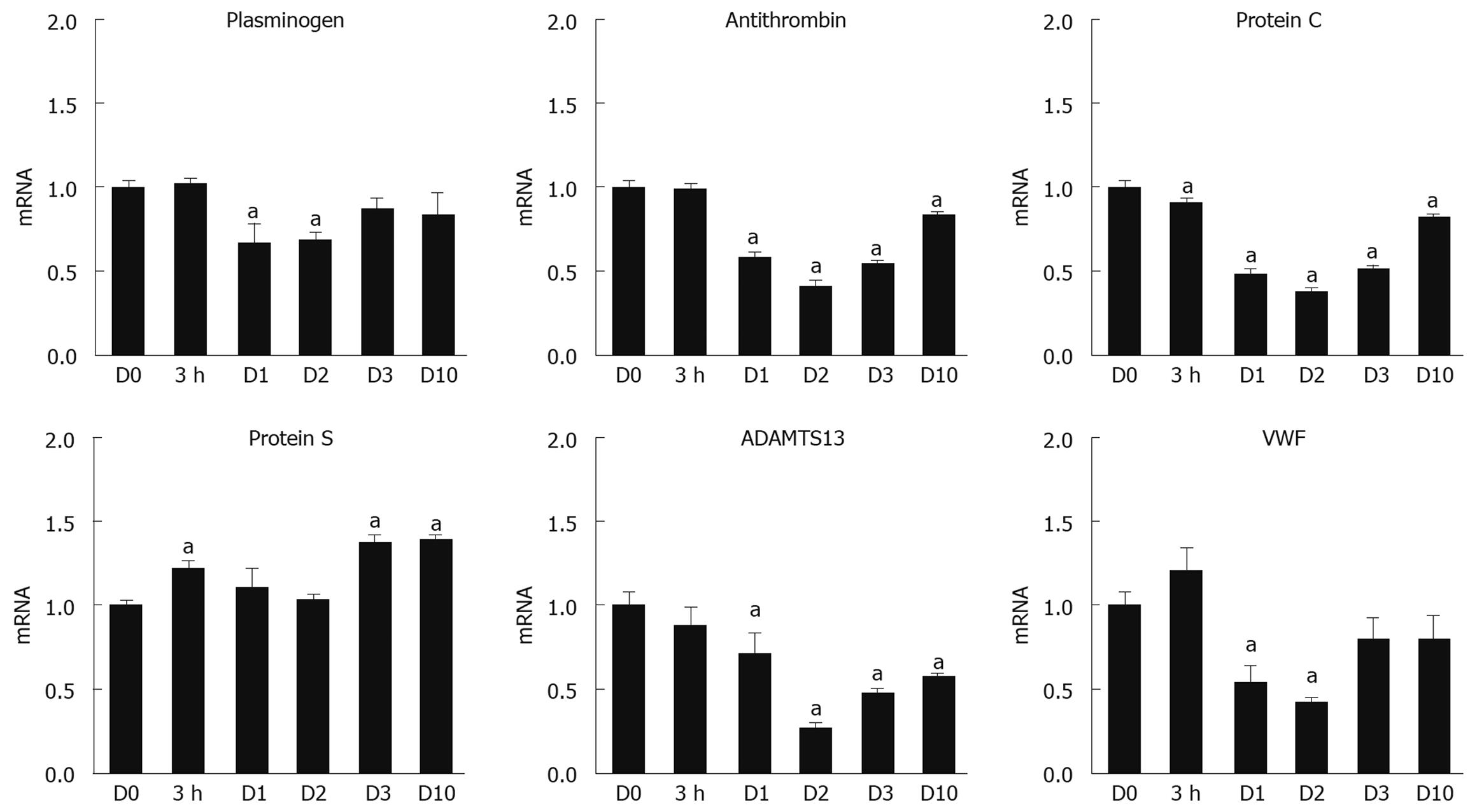
Figure 2 Gene expression profiling of coagulation-related factors in the liver during the DH-mediated regenerative phase following TCPOBOP induction.
The mRNA expression levels of 6 coagulation-related factors [plasminogen, antithrombin, protein C, protein S, a disintegrin-like and metalloproteinase with thrombospondin type 1 motif 13 (ADAMTS13), and von Willebrand factor (VWF)] were assessed in mouse livers at D0, 3 h, D1, D2, D3, and D10 by real-time RT-PCR (n = 8/time point). The mRNA levels of each gene were normalized to the geometric mean of PPIA and RPL4 mRNA levels. The values were expressed as a comparative ratio to the D0 samples, and represented as the mean ± SE. aP < 0.05 vs D0.
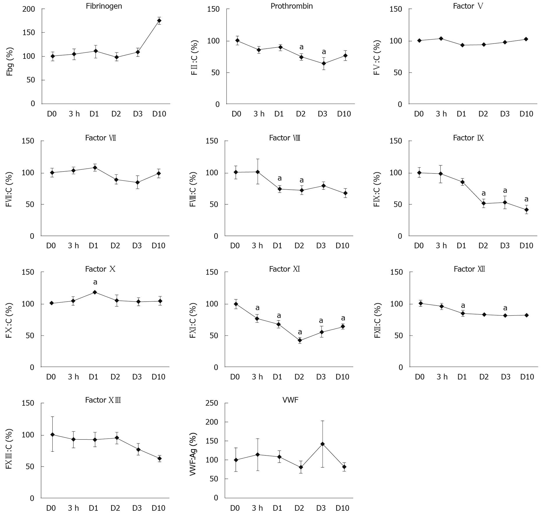
Figure 3 Determination of plasma levels and activity for coagulation factors during the DH-mediated regenerative phase following TCPOBOP induction.
Plasma activity levels of 10 coagulation factors (fibrinogen, prothrombin, factor V, VII, VIII, IX, X, XI, XII, and XIII) and plasma von Willebrand factor (VWF) antigen levels were assessed in mice during the DH-mediated regenerative phase. Activity levels of prothrombin, factor V, VII, VIII, IX, X, XI, and XII were measured by one-stage clotting assay, fibrinogen activities were measured by the Clauss method, factor XIII activities were measured by chromogenic assay, and VWF antigen levels were measured by specific ELISA. The plasma samples were obtained at D0, 3 h, D1, D2, D3, and D10 after TCPOBOP injection (n = 8/time point). The data were described as percentage of pooled normal plasma, and represented as the mean ± SE. aP < 0.05 vs D0.
- Citation: Tatsumi K, Ohashi K, Taminishi S, Takagi S, Utoh R, Yoshioka A, Shima M, Okano T. Effects on coagulation factor production following primary hepatomitogen-induced direct hyperplasia. World J Gastroenterol 2009; 15(42): 5307-5315
- URL: https://www.wjgnet.com/1007-9327/full/v15/i42/5307.htm
- DOI: https://dx.doi.org/10.3748/wjg.15.5307


