©2009 The WJG Press and Baishideng.
World J Gastroenterol. Jan 28, 2009; 15(4): 489-495
Published online Jan 28, 2009. doi: 10.3748/wjg.15.489
Published online Jan 28, 2009. doi: 10.3748/wjg.15.489
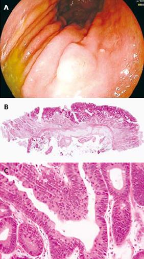
Figure 1 A case of true HGIEN of gastric mucosa.
A: Endoscopic view of a sessile polypoid lesion (types 0-I) in the greater curvature of gastric corpus. The lesion is approximately 1.2 cm in size with a smooth contour. Biopsy pathology indicated high grade intraepithelial neoplasia; B: Low-power view of the en-bloc resected specimen, showing negative vertical and horizontal margins (HE staining, × 25); C: High-power view shows prominent cellular atypia and increased proliferative activity without stromal invasion, indicating high grade intraepithelial neoplasia (HE staining, × 200).
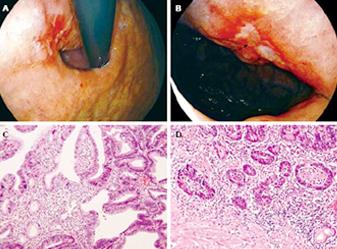
Figure 2 Presence of ulcer plaque is associated with gastric cancer.
A: Retroflex view of a flat depressed lesion in the lesser curvature of cardia. The lesion is presented with a reddish area approximately 1.4 cm in size, scattered with irregular ulcer plaque; B: Forward view of the same lesion. The ulcer plaque can be clearly observed with a size larger than 5 mm (arrows); C: Low-power view of biopsy specimen shows irregular tubules with increased branching and architectural distortion. Prominent cellular atypia can be noted, indicating high grade intraepithelial neoplasia (HE staining, × 100); D: The entire lesion was removed by surgery which showed tumor invasion into lamina propria and partly the muscularis mucosae (HE staining, × 100). The final diagnosis was a type 0-IIc well-differentiated adenocarcinoma with muscularis mucosae invasion, T1 N0 M0.
- Citation: Wu W, Wu YL, Zhu YB, Wei Q, Guo Y, Zhu ZG, Yuan YZ. Endoscopic features predictive of gastric cancer in superficial lesions with biopsy-proven high grade intraepithelial neoplasia. World J Gastroenterol 2009; 15(4): 489-495
- URL: https://www.wjgnet.com/1007-9327/full/v15/i4/489.htm
- DOI: https://dx.doi.org/10.3748/wjg.15.489


