©2009 The WJG Press and Baishideng.
World J Gastroenterol. Jan 28, 2009; 15(4): 478-483
Published online Jan 28, 2009. doi: 10.3748/wjg.15.478
Published online Jan 28, 2009. doi: 10.3748/wjg.15.478
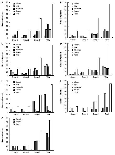
Figure 1 Results of analysis of histologic features present in intrahepatic neonatal cholestasis (IHNC).
A: Cholestasis: there was no significant difference between the 3 groups studied (P > 0.05); B: Giant cells: there was no significant difference between the 3 groups studied (P > 0.05); C: Eosinophils: there was no significant difference between the 3 groups studied (P > 0.05); D: Erythropoiesis: there was a significant difference in group 1 (P < 0.05); E: Siderosis: there was no significant difference between the 3 groups studied (P > 0.05); F: Portal fibrosis (graded as absent, mild, moderate, and severe): there was no significant difference between the 3 groups studied (P > 0.05); G: Septum (graded as absent or present) in groups 1 (infectious), 2 (genetic-endocrine-metabolic) and 3 (idiopathic): there was no significant difference between the 3 groups studied (P > 0.05).
- Citation: Bellomo-Brandao MA, Escanhoela CA, Meirelles LR, Porta G, Hessel G. Analysis of the histologic features in the differential diagnosis of intrahepatic neonatal cholestasis. World J Gastroenterol 2009; 15(4): 478-483
- URL: https://www.wjgnet.com/1007-9327/full/v15/i4/478.htm
- DOI: https://dx.doi.org/10.3748/wjg.15.478


