©2009 The WJG Press and Baishideng.
World J Gastroenterol. Jan 28, 2009; 15(4): 412-416
Published online Jan 28, 2009. doi: 10.3748/wjg.15.412
Published online Jan 28, 2009. doi: 10.3748/wjg.15.412
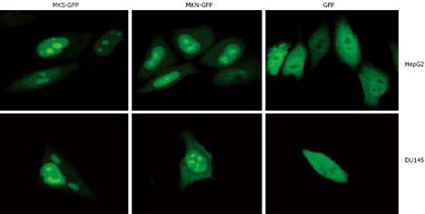
Figure 1 Subcellular localization analysis of MKS-GFP and MKN-GFP fusion proteins in carcinoma cell lines.
MKS-GFP and MKN-GFP fusion proteins were localized to nuclei and accumulated in nucleoli, but rarely localized to cytoplasm. The high light “spots” in the center of nuclei are nucleoli. HepG2 cells (upper panel) and DU145 cells (bottom panel) are transfected with pEGFP-N2, and diffuse freely in the whole cell except in nucleoli.
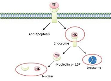
Figure 2 Nuclear targeting by growth factor MK.
MK was endocytosed into cell cytosol through binding to the receptor of LRP, and then tanslocated to nuclei in the presence of nucleulin or LBP. At the same time, the internalized MK is degraded by proteasome to “off” the signals from the cell surface receptors.
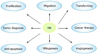
Figure 3 Biological function and medical application of MK.
The most attractive feature of MK is its involvement in carcinogenesis, which plays a great role in proliferation, migration, anti-apoptosis, mitogenesis, transformation, and angiogenesis. Furthermore, MK is expected to be a target molecule of diagnosis of and therapy for tumor.
- Citation: Dai LC. Midkine translocated to nucleoli and involved in carcinogenesis. World J Gastroenterol 2009; 15(4): 412-416
- URL: https://www.wjgnet.com/1007-9327/full/v15/i4/412.htm
- DOI: https://dx.doi.org/10.3748/wjg.15.412


