©2009 The WJG Press and Baishideng.
World J Gastroenterol. Jan 28, 2009; 15(4): 407-411
Published online Jan 28, 2009. doi: 10.3748/wjg.15.407
Published online Jan 28, 2009. doi: 10.3748/wjg.15.407
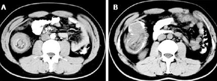
Figure 1 Abdominal computed tomography in adult intussusception.
A: The characteristic “target”-shaped soft-tissue mass with a layering effect of a 29-year old male patient with a diffuse, small B-cell (Burkitt-like) non Hodgkin lymphoma of the ileum who developed an ileo-colic intussusception; B: A “sausage”-shaped soft tissue mass in the ascending colon of the same patient.
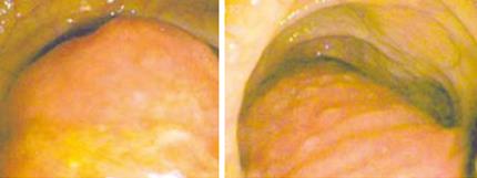
Figure 2 Colonoscopy.
Revealing the presence of the inverted terminal ileum (intussusceptum) in the ascending colon (intussuscipiens) in a patient with an ileo-cecal intussusception due to an ileal lipoma.
Figure 3 Intraoperative findings.
A: Thickened, congested and inflamed terminal ileum with proximal small bowel obstruction in a 75-year old woman with ileo-colonic intussusception; B: The surgical specimen after the en bloc resection of the terminal ileum and the ascending colon in the same patient; C: The cause of the intussusception was a lipoma of the ileo-cecal valve (arrow).
- Citation: Marinis A, Yiallourou A, Samanides L, Dafnios N, Anastasopoulos G, Vassiliou I, Theodosopoulos T. Intussusception of the bowel in adults: A review. World J Gastroenterol 2009; 15(4): 407-411
- URL: https://www.wjgnet.com/1007-9327/full/v15/i4/407.htm
- DOI: https://dx.doi.org/10.3748/wjg.15.407


