©2009 The WJG Press and Baishideng.
World J Gastroenterol. Sep 14, 2009; 15(34): 4240-4262
Published online Sep 14, 2009. doi: 10.3748/wjg.15.4240
Published online Sep 14, 2009. doi: 10.3748/wjg.15.4240
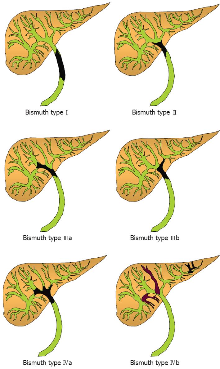
Figure 1 Bismuth’s classification of cholangiocarcinomas.
Type I: Cholangiocarcinoma is confined to the common hepatic duct; Type II: Cholangiocarcinoma involves the common hepatic duct bifurcation; Type IIIa: Cholangiocarcinoma affects the hepatic duct bifurcation and the right hepatic duct; Type IIIb: Cholangiocarcinoma affects the hepatic duct bifurcation and the left hepatic duct; Type IV: Cholangiocarcinoma is either located at the biliary confluence with both the right and left hepatic ducts involvement or has multifocal distribution.
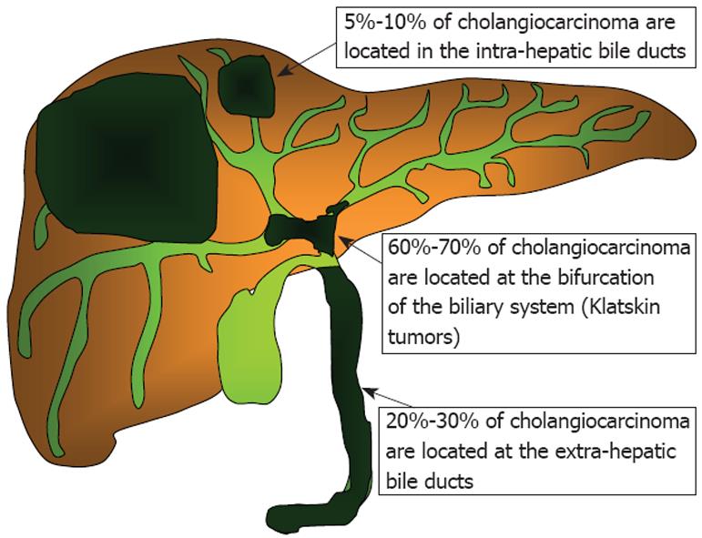
Figure 2 Anatomical presentation of cholangiocarcinomas.
The majority of cholangiocarcinomas (60%-70%) present in the area of the biliary duct bifurcation and are called Klatskin tumors. The extra-hepatic bile duct is involved in 20%-30% of cases while intrahepatic cholangiocarcinomas represent 5%-10% of the tumors originating from the biliary system.
- Citation: Aljiffry M, Walsh MJ, Molinari M. Advances in diagnosis, treatment and palliation of cholangiocarcinoma: 1990-2009. World J Gastroenterol 2009; 15(34): 4240-4262
- URL: https://www.wjgnet.com/1007-9327/full/v15/i34/4240.htm
- DOI: https://dx.doi.org/10.3748/wjg.15.4240


