©2009 The WJG Press and Baishideng.
World J Gastroenterol. Aug 28, 2009; 15(32): 4037-4043
Published online Aug 28, 2009. doi: 10.3748/wjg.15.4037
Published online Aug 28, 2009. doi: 10.3748/wjg.15.4037
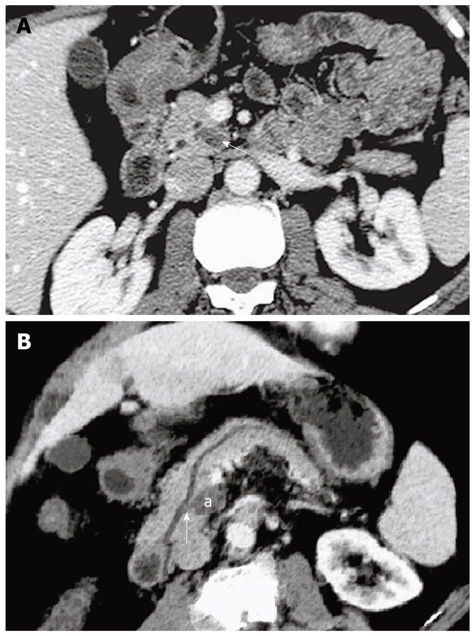
Figure 1 Pathologically confirmed benign branch-duct-type IPMN in a 47-year-old woman with abdominal discomfort for about 6 mo.
A: There was a 2-cm cystic mass (white arrow) in the uncinate process of the pancreas at the axial abdominal MDCT image; B: The cystic mass (a) in the posterior pancreatic parenchyma was demonstrated with a communication (white arrow) between the mass and the main pancreatic duct which was slightly dilated in the curved reformed (CR) image.
Figure 2 Pathologically confirmed malignant combined-type IPMN in a 41-year-old man with abdominal and back pain for about 2 years.
A: A 4 cm cystic mass with multiple septa arising from the pancreatic head was revealed in the axial MDCT image; B: Besides the cystic mass in the pancreatic head, the profile of the main pancreatic duct, which was severely dilated, was depicted on the CR image; C: The cystic lesion (a) in the pancreatic head invaded the duodenum and main portal vein resulting in the duodenal wall thickening (white arrow) with marked enhancement and irregular narrowing (dark arrow) of the vessel in the multiplanar volume reformation (MPVR) image.
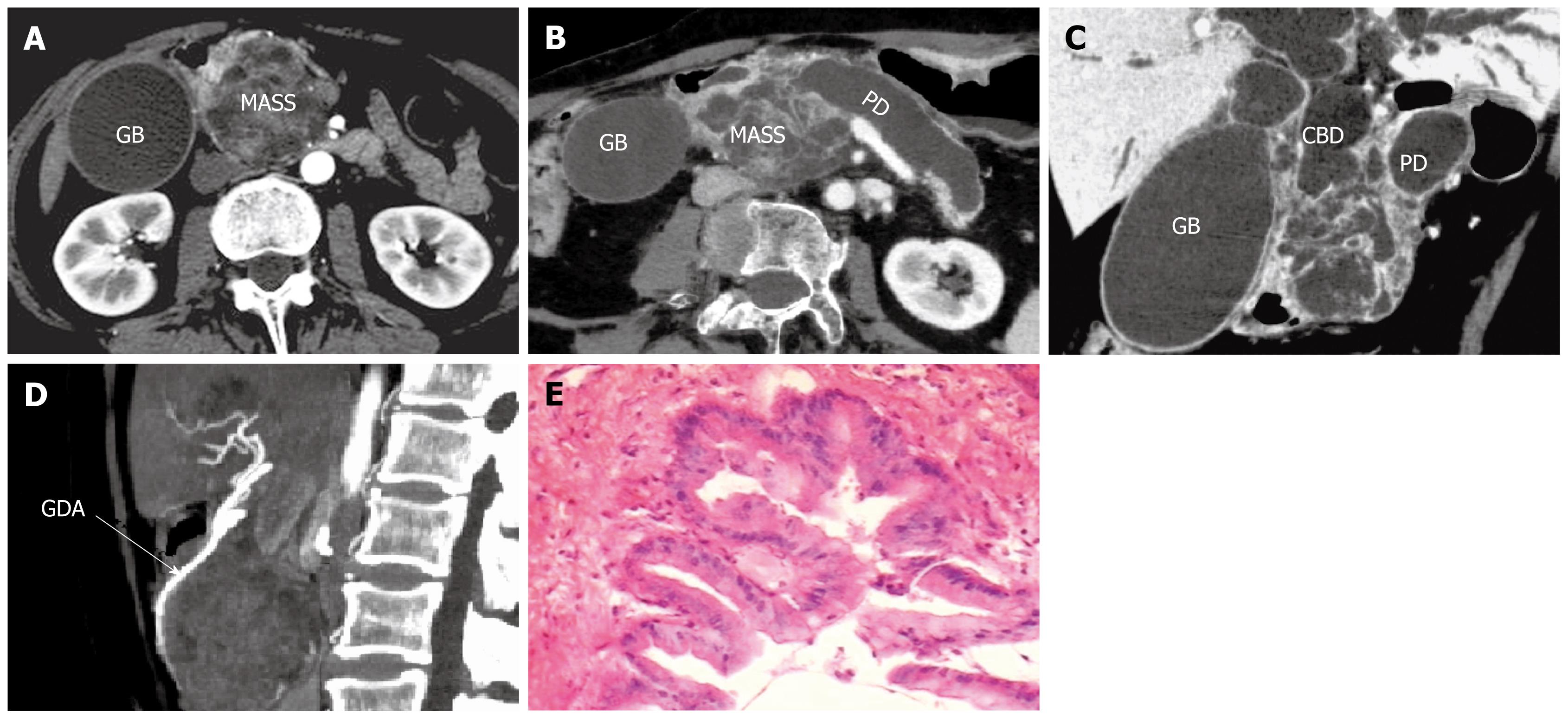
Figure 3 Pathologically confirmed malignant combined-type IPMN in a 65-year-old man with jaundice and abdominal pain for about 1 year.
A: An 8 cm cystic and solid mass (MASS) was seen in the axial arterio-phased MDCT image with contrast agents. The gallbladder (GB) was distended; B: The heterogeneous mass was shown with severe dilatation of the main pancreatic duct (PD) and the gallbladder (GB) in the CR image; C: The profile of the dilatation of pancreatobiliary system (CBD, common bile duct) was entirely depicted in the MPVR image; D: The gastroduodenal artery (GDA) showed irregularity as a result of infiltration of the tumor; E: The tumor consisted of papillary proliferations of tall columnar mucin-producing epithelium. Atypical epithelium characterized by enlarged nuclei (HE, × 150).
Figure 4 Pathologically confirmed malignant combined-type IPMN in a 55-year-old man with abdominal pain for 1.
5 years. A: A 3 cm × 10 cm longitudinally cystic mass in the pancreatic body was shown with a mural nodule (white arrow) in the axial MDCT images; B: The classification of combined type for this case was accurately defined by the CR image; C: The profile of the cystic mass (a) and dilatation of the branch duct and the upstream pancreatic duct were identified in the MPVR images.
- Citation: Tan L, Zhao YE, Wang DB, Wang QB, Hu J, Chen KM, Deng XX. Imaging features of intraductal papillary mucinous neoplasms of the pancreas in multi-detector row computed tomography. World J Gastroenterol 2009; 15(32): 4037-4043
- URL: https://www.wjgnet.com/1007-9327/full/v15/i32/4037.htm
- DOI: https://dx.doi.org/10.3748/wjg.15.4037


