©2009 The WJG Press and Baishideng.
World J Gastroenterol. Aug 21, 2009; 15(31): 3940-3943
Published online Aug 21, 2009. doi: 10.3748/wjg.15.3940
Published online Aug 21, 2009. doi: 10.3748/wjg.15.3940
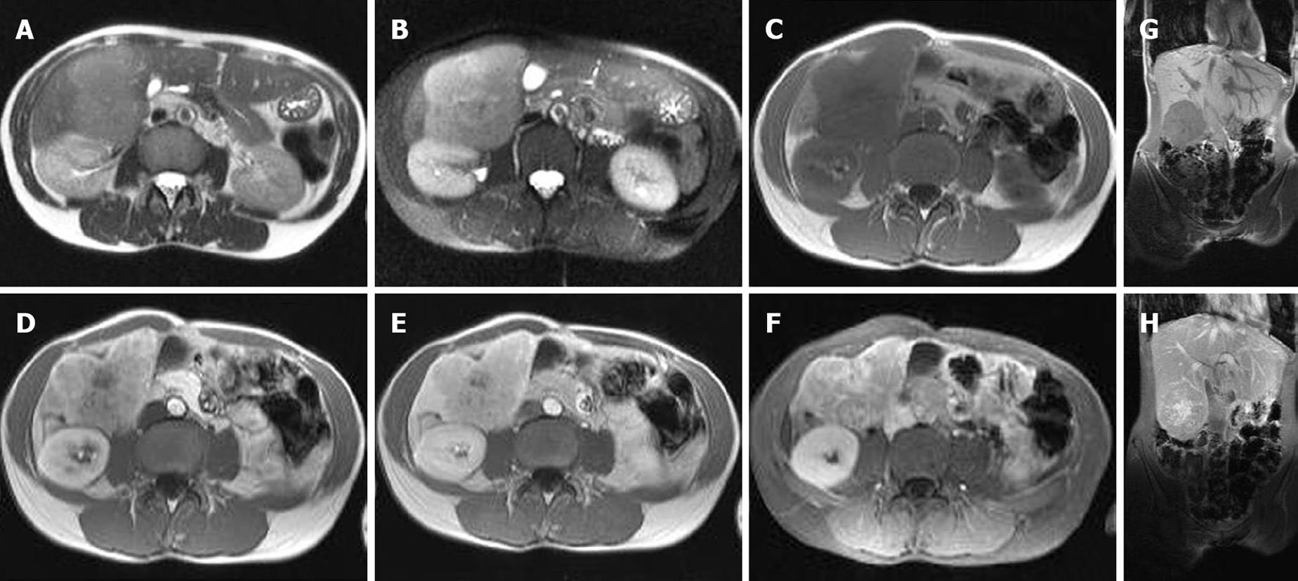
Figure 1 MRI images of combined hepatocellular and cholangiocellular carcinoma.
Axial T2 (A) and T2 fat saturated (B) MRI images showed a slightly well circumscribed lesion. The mass is hyperintense in comparison to the normal liver. Axial T1 (C) MRI images demonstrated a hypointense lesion. Early arterial (D), late arterial (E) and venous (F) MRI images displayed a hypervascular heterogeneous enhancement with central scar. The coronal T1 (G) image showed a hypointense lesion, while the coronal T1 MRI image after Gadolinium contrast (H) demonstrated a heterogeneous early enhancement of the lesion with delayed uptake in the scar.
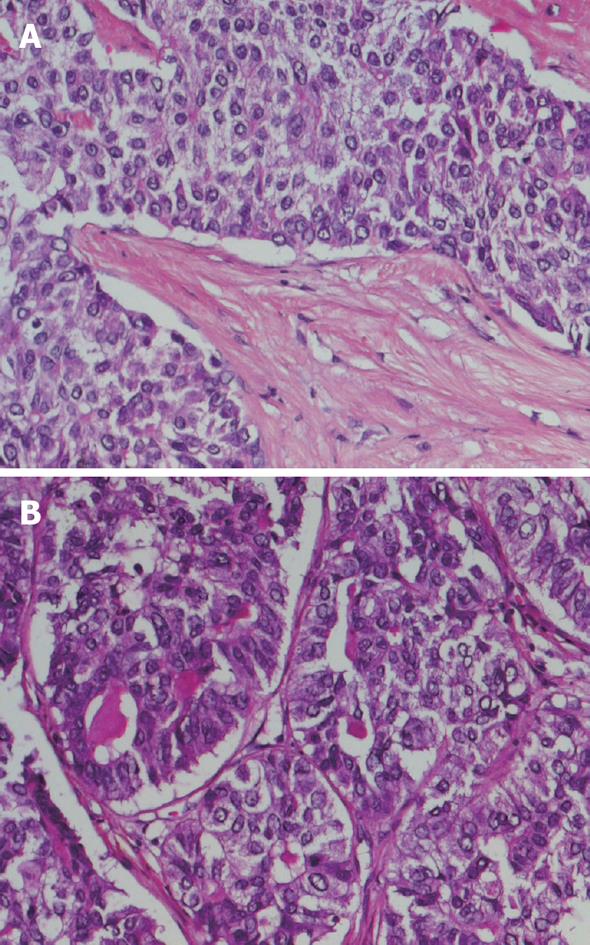
Figure 2 Histopathological images of combined hepatocellular and cholangiocellular carcinoma.
A: Nests of tumor cells with trabecular architecture, reminiscent of hepatocellular differentiation, separated by bands of fibrous tissue; B: Focally tumor cells formed glandular structures with mucinous secretory material, compatible with cholangiocellular differentiation.
- Citation: Willekens I, Hoorens A, Geers C, Op de Beeck B, Vandenbroucke F, de Mey J. Combined hepatocellular and cholangiocellular carcinoma presenting with radiological characteristics of focal nodular hyperplasia. World J Gastroenterol 2009; 15(31): 3940-3943
- URL: https://www.wjgnet.com/1007-9327/full/v15/i31/3940.htm
- DOI: https://dx.doi.org/10.3748/wjg.15.3940


