Copyright
©2009 The WJG Press and Baishideng.
World J Gastroenterol. Jul 28, 2009; 15(28): 3576-3579
Published online Jul 28, 2009. doi: 10.3748/wjg.15.3576
Published online Jul 28, 2009. doi: 10.3748/wjg.15.3576
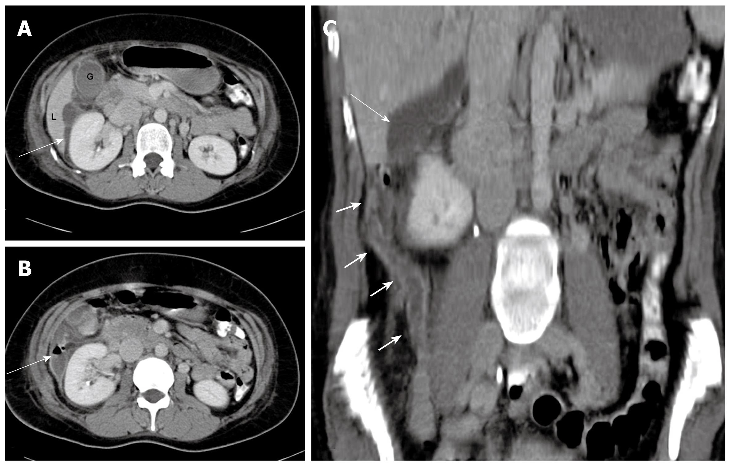
Figure 1 A 30-year-old woman presenting with a clinical diagnosis of acute cholecystitis.
A and B: Contrast-enhanced computed tomography (CT) sections showing inflammatory changes (arrow) adjacent to the inferior tip of the liver (L); B: Thickened appendix (arrow) with mild inflammatory changes in the retrocecal region; C: Coronal reconstruction showing the extent of inflammatory changes (arrows) from the retrocecal region to the tip of the liver.
Figure 2 A 31-year-old woman presenting with right hypochondrial pain and a clinical diagnosis of pelvic inflammatory disease and right pyelonephritis.
A: Contrast-enhanced CT scan showing fluid collection (arrow) in the subhepatic region, extending anteriorly to the gallbladder fossa with inflammatory stranding; B: Note the air fluid level in the collection adjacent to the right kidney; C: Coronal reconstruction showing the long thickened and inflamed appendix (short arrows) reaching the subhepatic region, and the subhepatic collection (arrow) is seen extending more cranially.
Figure 3 A 34-year-old man with colicky right flank pain and clinical diagnosis of right ureteric colic.
A: Ultrasound showed a subhepatic fluid collection (arrows) and no other significant abnormality; B: CT scan performed 2 d later showed the collection in the subhepatic region (short arrow). Note the air-fluid level in the anterior collection (long arrow) with inflammatory changes; C: The section at the level of the cecum and appendix shows inflammatory changes in the retrocecal region (short arrow) and thickened appendix (long arrow).
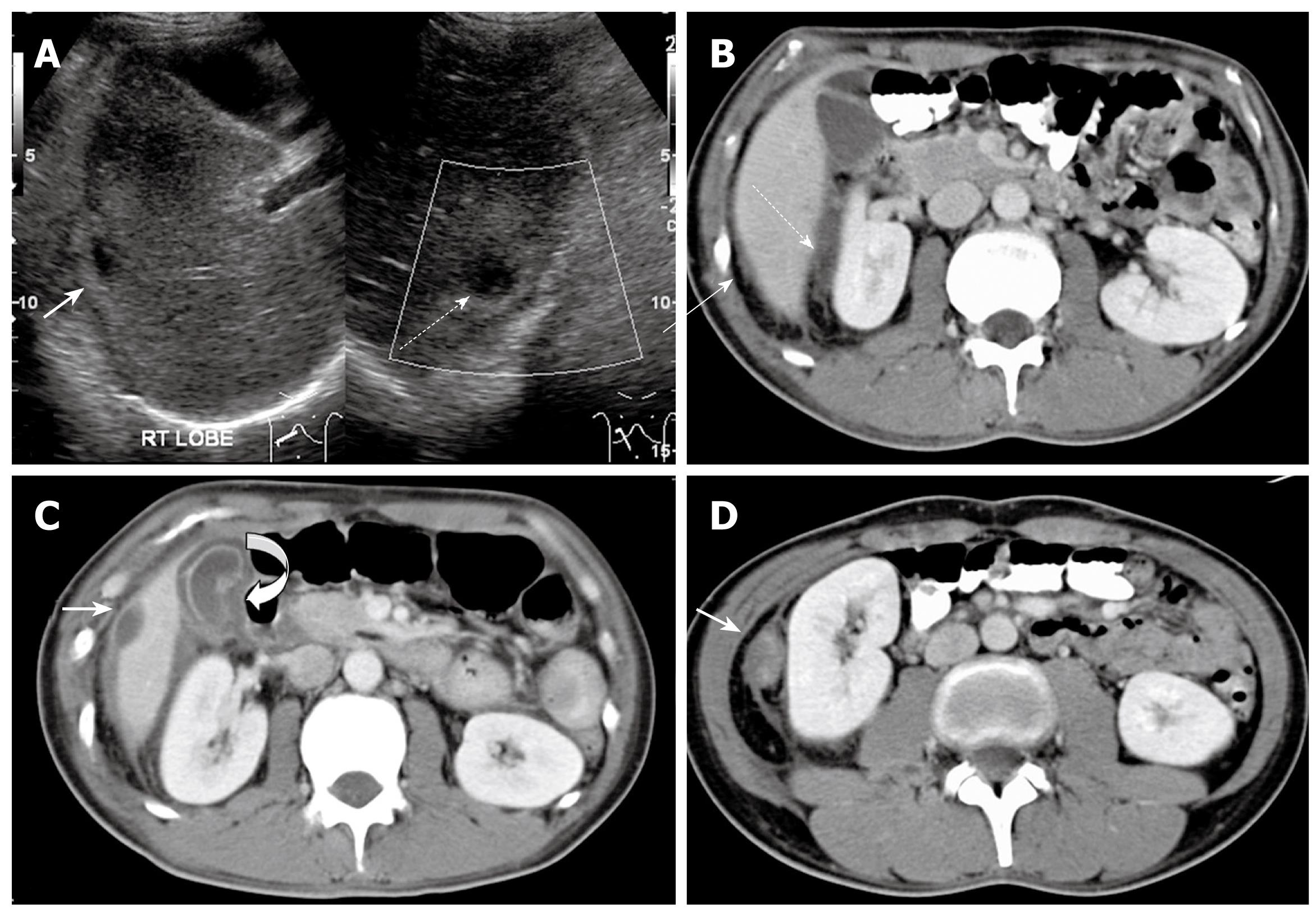
Figure 4 A 27-year-old man with recurrent right upper abdominal pain.
A: Ultrasound showed a hypoechoic area in the subphrenic (straight arrow) and subhepatic (broken arrow) region; B: Confirmation by contrast-enhanced CT; C: CT also showed a thickened gallbladder wall (curved arrow), subhepatic collection (white arrow) and inflammation in the perinephric region; D: Another caudal section shows a thickened appendix with inflammatory stranding in the perinephric region.
- Citation: Ong EMW, Venkatesh SK. Ascending retrocecal appendicitis presenting with right upper abdominal pain: Utility of computed tomography. World J Gastroenterol 2009; 15(28): 3576-3579
- URL: https://www.wjgnet.com/1007-9327/full/v15/i28/3576.htm
- DOI: https://dx.doi.org/10.3748/wjg.15.3576


