Copyright
©2009 The WJG Press and Baishideng.
World J Gastroenterol. Jul 28, 2009; 15(28): 3565-3568
Published online Jul 28, 2009. doi: 10.3748/wjg.15.3565
Published online Jul 28, 2009. doi: 10.3748/wjg.15.3565
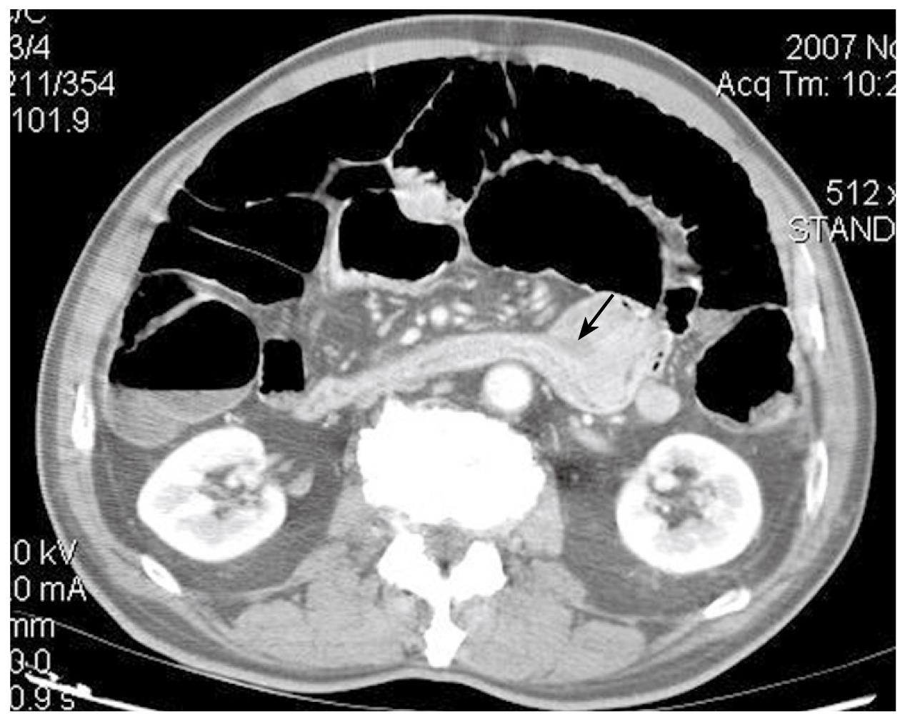
Figure 1 CT showing a mass on the fourth part of the duodenum (arrow).
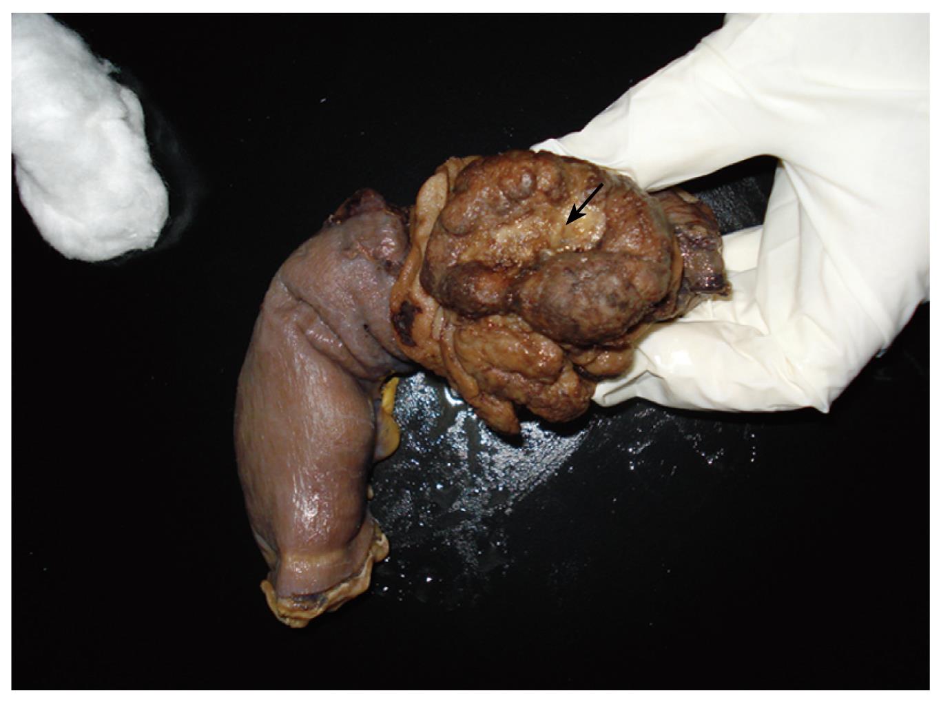
Figure 2 Gross appearance of the surgical specimen containing the tumor (arrow).
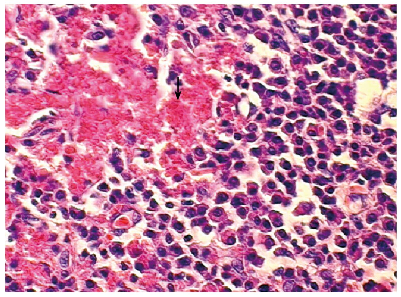
Figure 3 Histopathological examination displaying a dense and diffuse infiltrate of plasma cells and amyloid deposit (arrow).
(HE × 400).
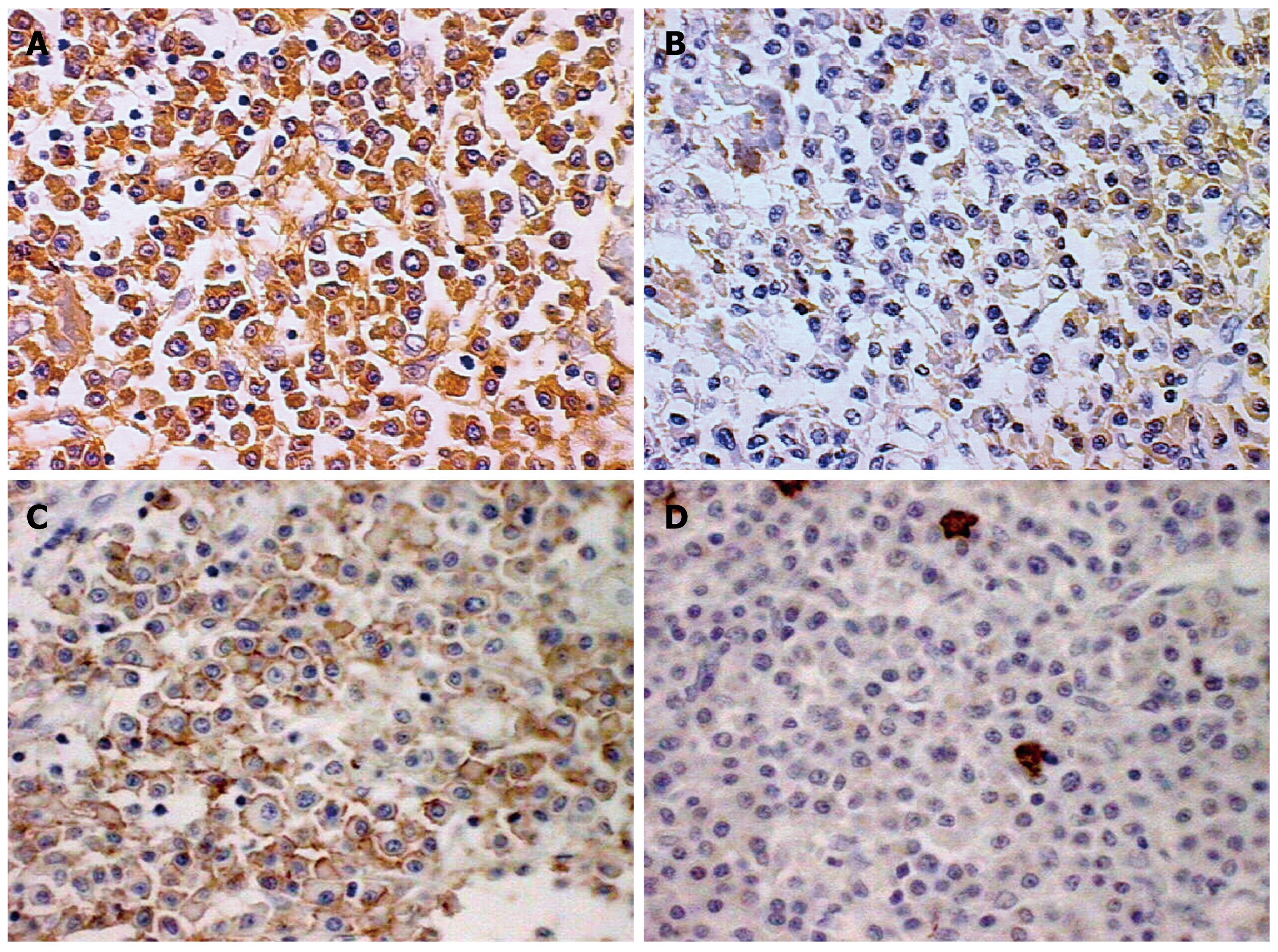
Figure 4 Immunohistochemical findings (positive cells in brown, × 400).
A: Most plasmocytoid cells were positive for κ light chain; B: Few plasmacytoid cells showing λ light chain staining; Plasmocytoid cells were positive for CD56 (C) and negative for CD20 (D).
- Citation: Carneiro FP, Sobreira MNM, Maia LB, Sartorelli AC, Franceschi LEAP, Brandão MB, Calaça BW, Lustosa FS, Lopes JV. Extramedullary plasmocytoma associated with a massive deposit of amyloid in the duodenum. World J Gastroenterol 2009; 15(28): 3565-3568
- URL: https://www.wjgnet.com/1007-9327/full/v15/i28/3565.htm
- DOI: https://dx.doi.org/10.3748/wjg.15.3565


