Copyright
©2009 The WJG Press and Baishideng.
World J Gastroenterol. Jul 28, 2009; 15(28): 3560-3564
Published online Jul 28, 2009. doi: 10.3748/wjg.15.3560
Published online Jul 28, 2009. doi: 10.3748/wjg.15.3560
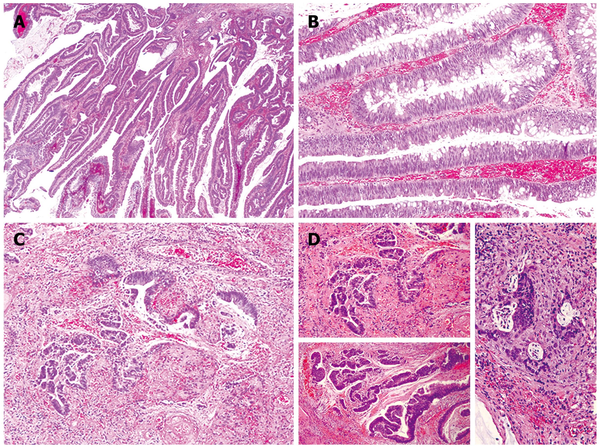
Figure 1 Microscopic sections.
A: The adenoma demonstrate villous architecture with long, delicate fronds (HE, × 40); B: The villous fronds are lined by dysplastic, intestinal-type epithelium with frequent goblet cells (HE, × 100); C and D: Multiple foci of invasive carcinoma are identified, with invasion confined to the lamina propria (HE, × 100).
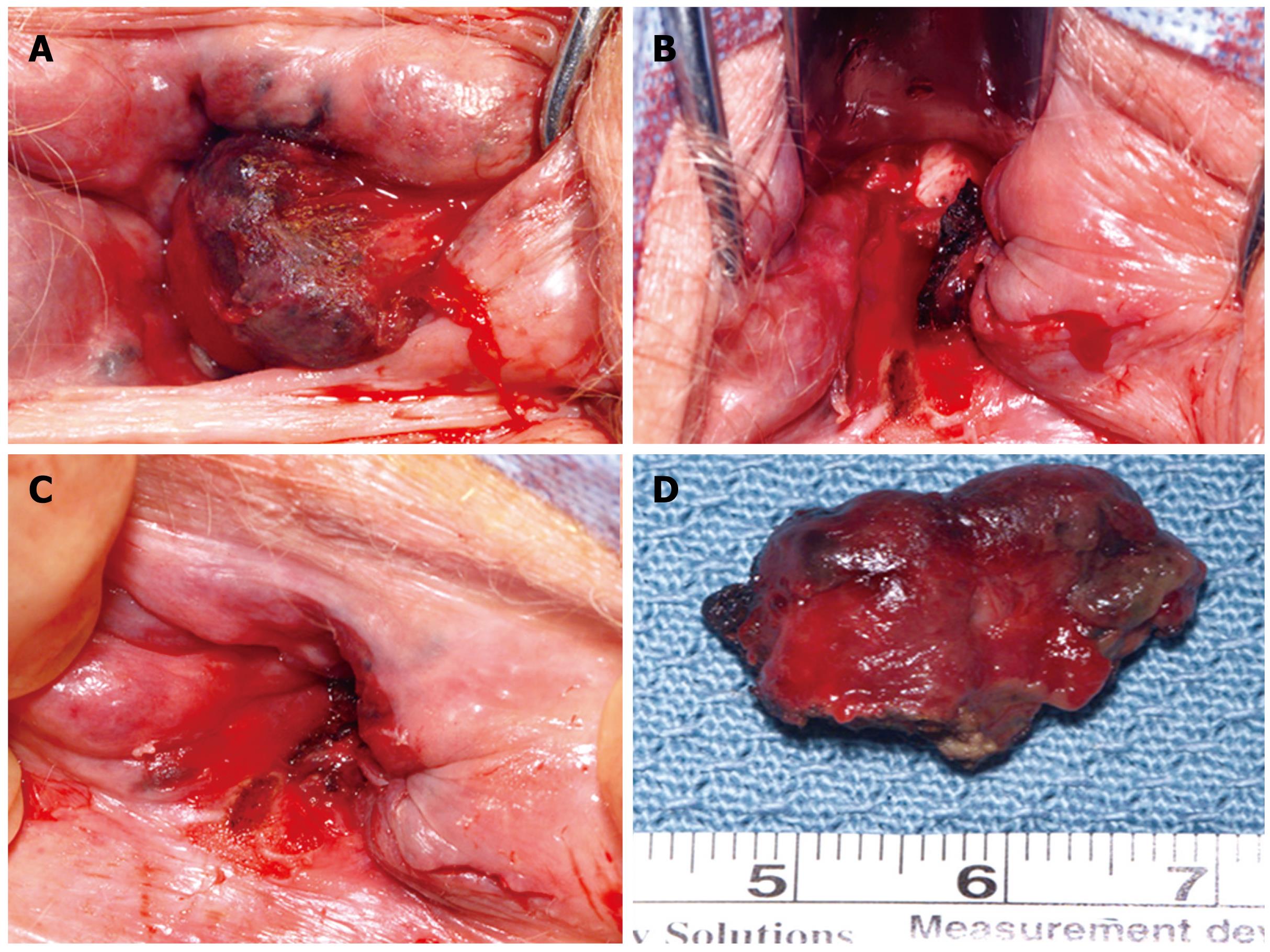
Figure 2 Intraoperative photographs.
A and D: Very large hemorrhoids up to 2 cm circumferentially located around the anal canal; B: The final appearance after complete excision of the hemorrhoids and the malignant mass; C: The area that contained the adenocarcinoma, previously excised in the emergency department, and prior to operating room excision.
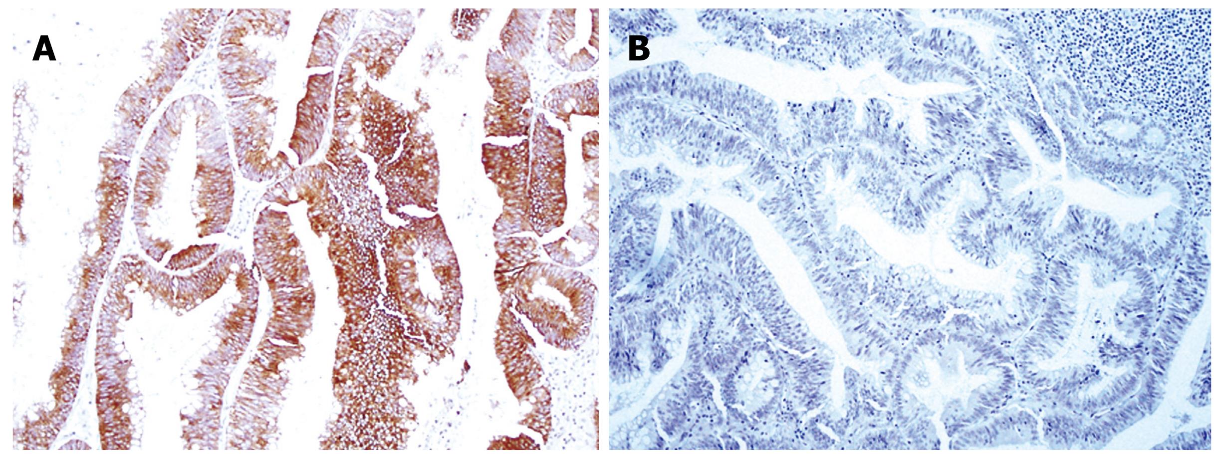
Figure 3 Immunohistochemical staining.
A: Cytokeratin 20 is diffusely positive within the epithelium (× 100); B: Cytokeratin 7 is negative (× 100).
- Citation: Colvin M, Delis A, Bracamonte E, Villar H, Jr LRL. Infiltrating adenocarcinoma arising in a villous adenoma of the anal canal. World J Gastroenterol 2009; 15(28): 3560-3564
- URL: https://www.wjgnet.com/1007-9327/full/v15/i28/3560.htm
- DOI: https://dx.doi.org/10.3748/wjg.15.3560


