Copyright
©2009 The WJG Press and Baishideng.
World J Gastroenterol. Jul 28, 2009; 15(28): 3511-3515
Published online Jul 28, 2009. doi: 10.3748/wjg.15.3511
Published online Jul 28, 2009. doi: 10.3748/wjg.15.3511
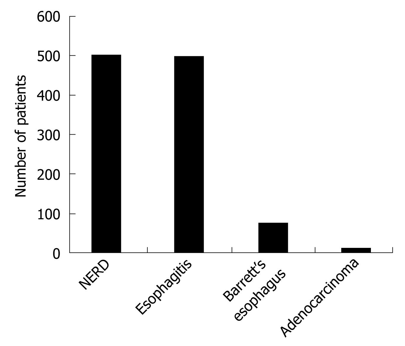
Figure 1 Endoscopic findings.
Esophagitis was detected in 498 patients (49.8%) and NERD was seen in 502 patients. BE was present in 73 patients. EAC was detected in eight patients.
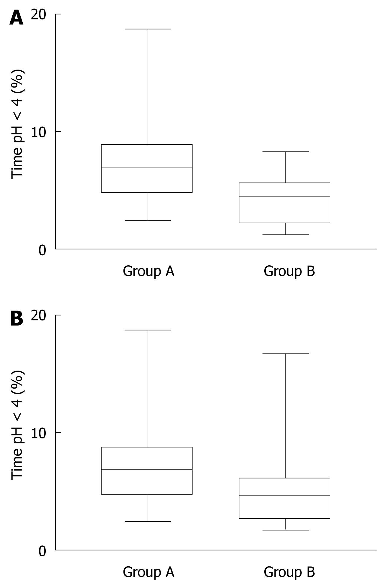
Figure 2 Distal esophageal acid exposure in both groups.
A: In the supine position. The median time at pH < 4 in the distal esophagus in the supine position was significantly longer in patients with BE (group A) than in those with GERD without BE (group B) (P < 0.03); B: In the upright position. The median time at pH < 4 in the distal esophagus in the upright position did not differ significantly between patients with BE (group A) and those with GERD without BE (group B).
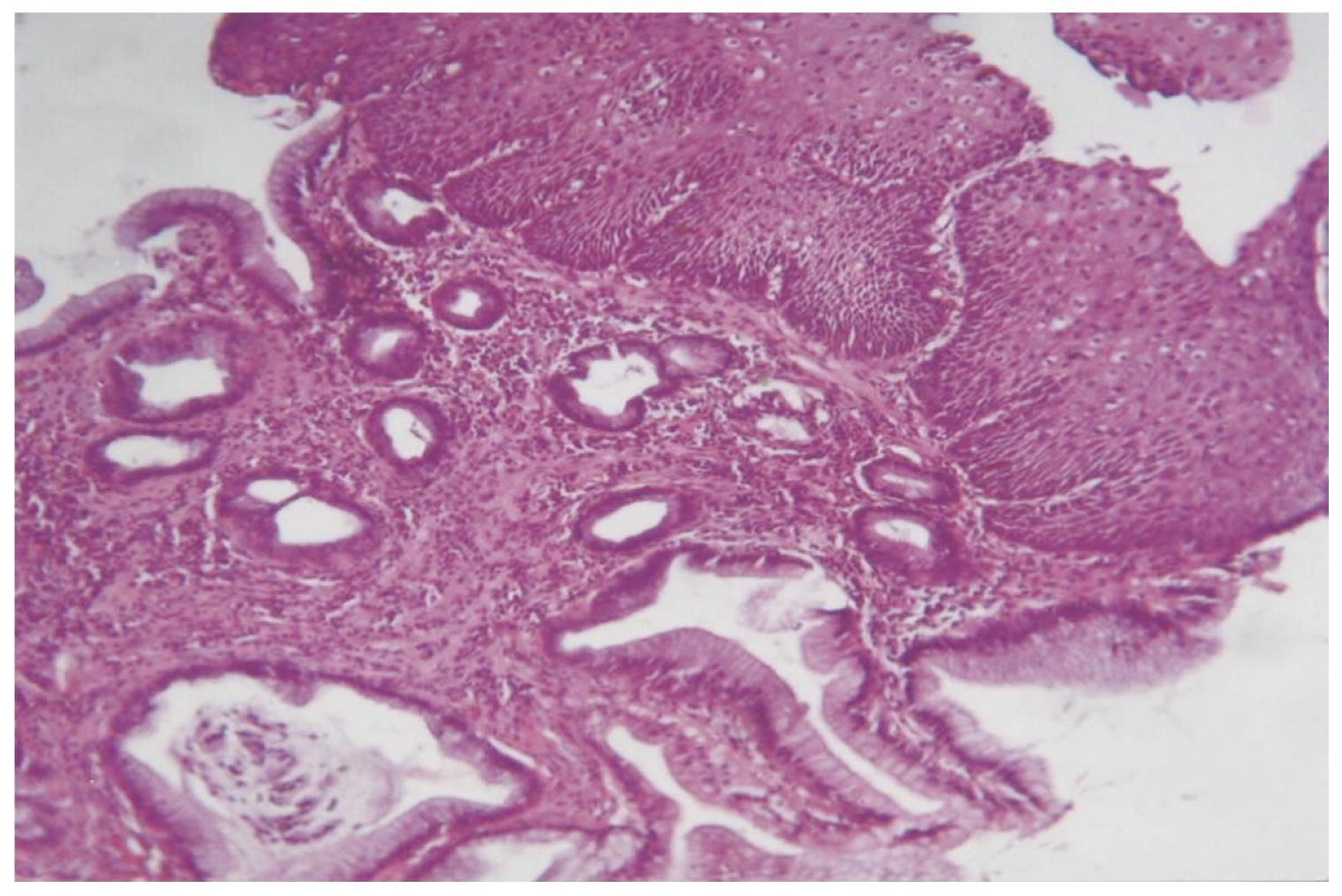
Figure 3 BE without dysplasia, showing true barrel-shaped goblet cells and intervening columnar cells, with incomplete brush-border, intestinal type metaplasia.
The squamous epithelium of the normal esophagus was transformed into columnar epithelium (HE, × 200).
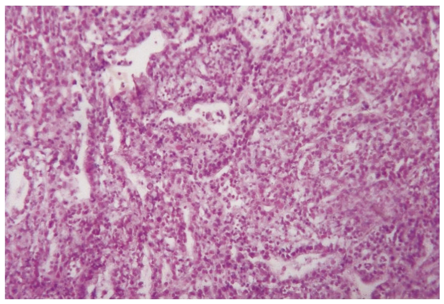
Figure 4 Well-differentiated adenocarcinoma showing gland formation, and low levels of nuclear pleomorphism and atypia (HE, × 200).
- Citation: Fouad YM, Makhlouf MM, Tawfik HM, Amin HE, Ghany WA, El-khayat HR. Barrett’s esophagus: Prevalence and risk factors in patients with chronic GERD in Upper Egypt. World J Gastroenterol 2009; 15(28): 3511-3515
- URL: https://www.wjgnet.com/1007-9327/full/v15/i28/3511.htm
- DOI: https://dx.doi.org/10.3748/wjg.15.3511


