Copyright
©2009 The WJG Press and Baishideng.
World J Gastroenterol. Jul 14, 2009; 15(26): 3319-3321
Published online Jul 14, 2009. doi: 10.3748/wjg.15.3319
Published online Jul 14, 2009. doi: 10.3748/wjg.15.3319
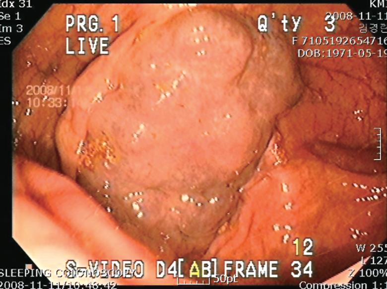
Figure 1 Colonoscopy of the cecum showing a bluish nodular lesion.
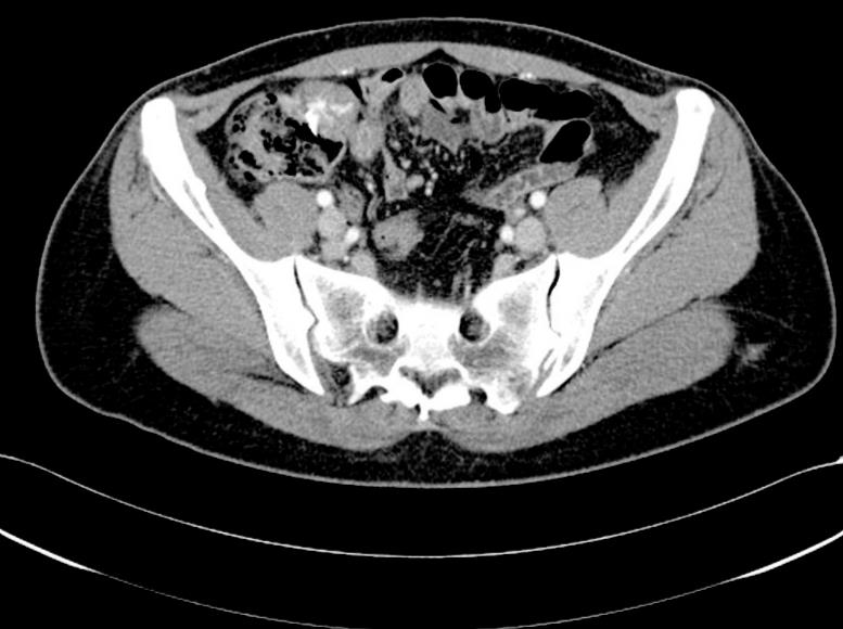
Figure 2 Abdominopelvic computed tomography showing a lesion in the cecum protruding extraluminally with heterogenous enhancement.
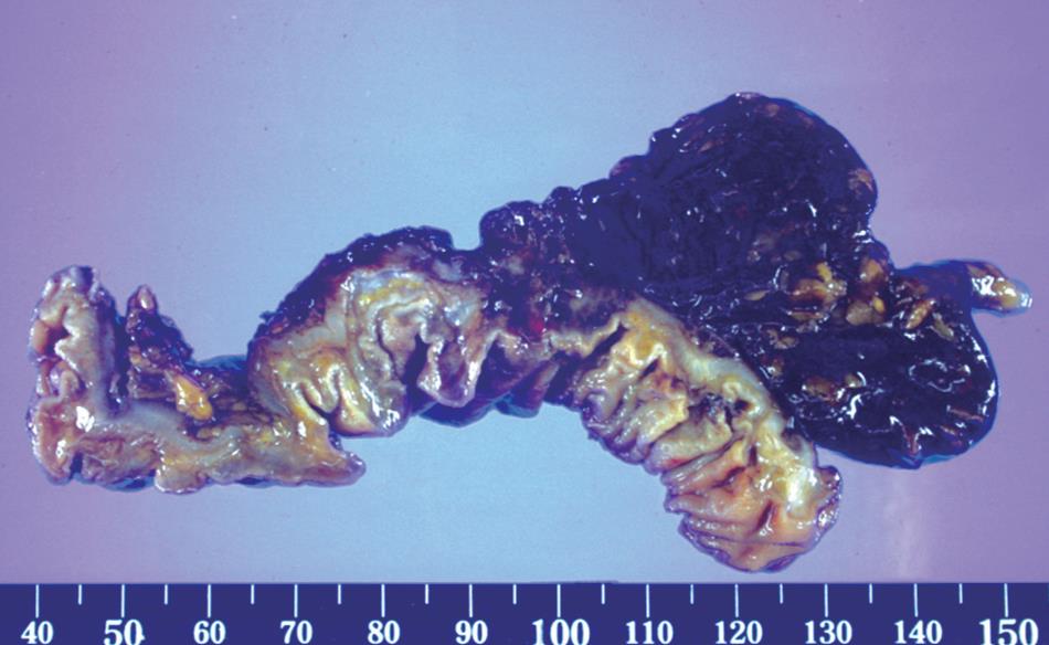
Figure 3 Macroscopically, the surgical specimen was a cavernous hemangioma.
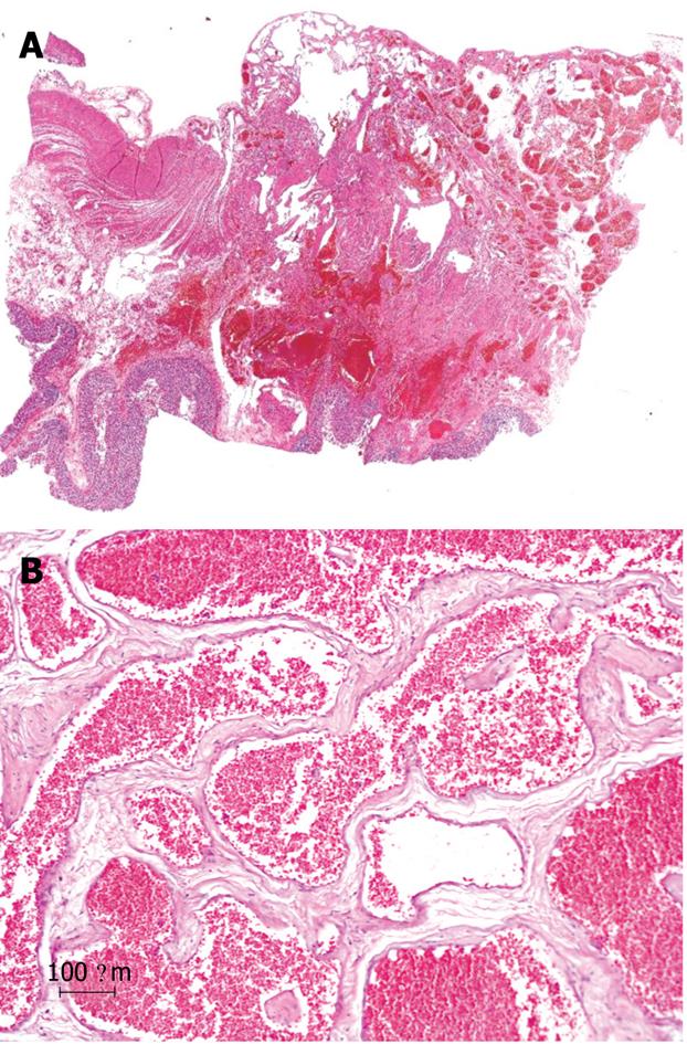
Figure 4 HE staining of the hemangioma.
A: Histological section showing large, dilated, blood-filled vessels lined by flattened endothelium (HE, × 1); B: The vascular walls were thickened focally by adventitial fibrosis (HE, × 100).
- Citation: Huh JW, Cho SH, Lee JH, Kim HR. Large cavernous hemangioma in the cecum treated by laparoscopic ileocecal resection. World J Gastroenterol 2009; 15(26): 3319-3321
- URL: https://www.wjgnet.com/1007-9327/full/v15/i26/3319.htm
- DOI: https://dx.doi.org/10.3748/wjg.15.3319


