Copyright
©2009 The WJG Press and Baishideng.
World J Gastroenterol. Jul 14, 2009; 15(26): 3283-3287
Published online Jul 14, 2009. doi: 10.3748/wjg.15.3283
Published online Jul 14, 2009. doi: 10.3748/wjg.15.3283
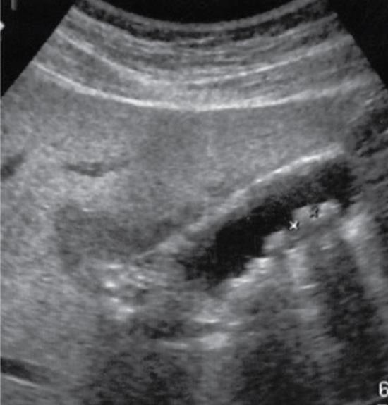
Figure 1 A 56-year-old man.
Several gallbladder stones measuring about 5-10 mm were detected by abdominal ultrasonography.
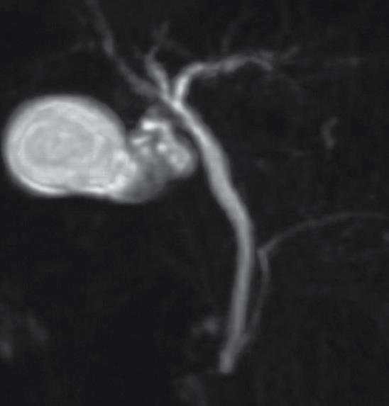
Figure 2 No translucency indicative of stones in the bile duct was shown by magnetic resonance cholangiopancreatography.
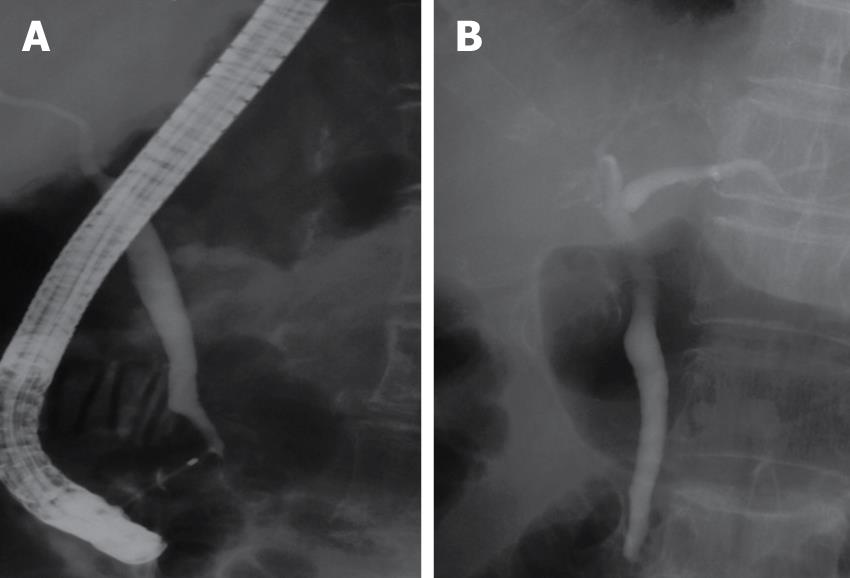
Figure 3 No translucency indicative of stones in the bile duct was shown by endoscopic retrograde cholangiopancreatography (A).
After withdrawal of the fiber, the posture was changed and the bile duct was imaged again. However, no translucency was observed (B).
Figure 4 Changes in serum.
A: Total-bilirubin; B: Alkaline phosphatase; C: Alanine transferase.
- Citation: Sakai Y, Tsuyuguchi T, Ishihara T, Yukisawa S, Ohara T, Tsuboi M, Ooka Y, Kato K, Katsuura K, Kimura M, Takahashi M, Nemoto K, Miyazaki M, Yokosuka O. Is ERCP really necessary in case of suspected spontaneous passage of bile duct stones? World J Gastroenterol 2009; 15(26): 3283-3287
- URL: https://www.wjgnet.com/1007-9327/full/v15/i26/3283.htm
- DOI: https://dx.doi.org/10.3748/wjg.15.3283


