©2009 The WJG Press and Baishideng.
World J Gastroenterol. Jul 14, 2009; 15(26): 3217-3227
Published online Jul 14, 2009. doi: 10.3748/wjg.15.3217
Published online Jul 14, 2009. doi: 10.3748/wjg.15.3217
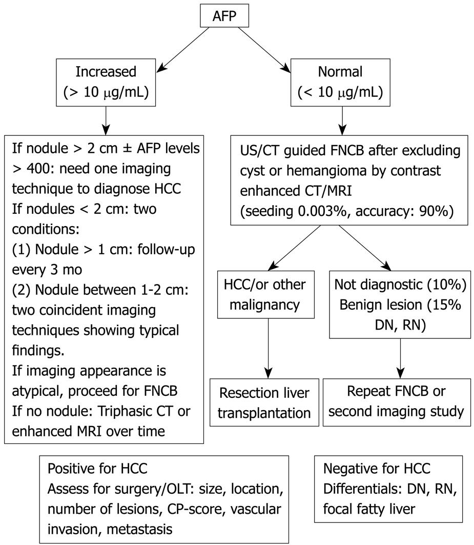
Figure 1 Algorithm for the investigation of a liver mass in a cirrhotic liver.
Some hepatologists consider biopsy to be unnecessary for a mass in a cirrhotic liver even if the α-fetoprotein (AFP) < 10; FNCB: Fine needle core biopsy; MRI: Magnetic resonance imaging.
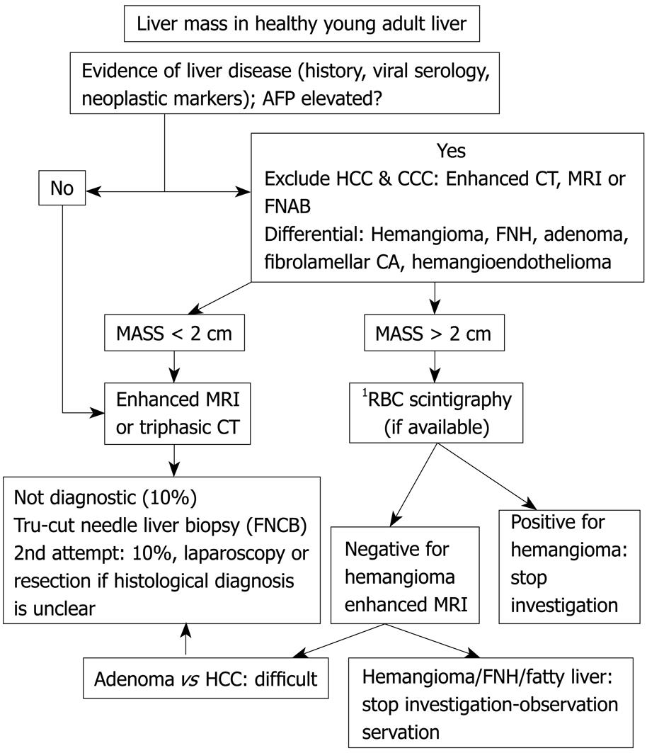
Figure 2 Algorithm for the management of a liver mass in a non-cirrhotic liver.
1Most centers do not use RBC scintigraphy to diagnose hemangioma due to their use of cross sectional imaging such as contrast enhanced ultrasonography (US)/CT/MRI.
- Citation: Assy N, Nasser G, Djibre A, Beniashvili Z, Elias S, Zidan J. Characteristics of common solid liver lesions and recommendations for diagnostic workup. World J Gastroenterol 2009; 15(26): 3217-3227
- URL: https://www.wjgnet.com/1007-9327/full/v15/i26/3217.htm
- DOI: https://dx.doi.org/10.3748/wjg.15.3217


