©2009 The WJG Press and Baishideng.
World J Gastroenterol. Jun 14, 2009; 15(22): 2809-2811
Published online Jun 14, 2009. doi: 10.3748/wjg.15.2809
Published online Jun 14, 2009. doi: 10.3748/wjg.15.2809
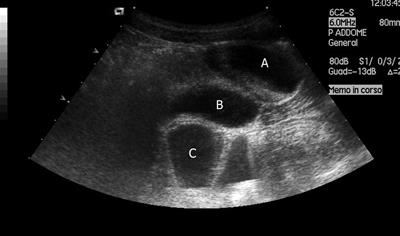
Figure 1 Abdominal ultrasound: anechoic oval supravesical lesion with fluid-fluid level (A); bladder (B); bowel (C).
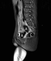
Figure 2 Abdominal MRI; T1 weighted sequence with fat-suppression (SPIR) after contrast medium, saggital plane: hypointense oval supravesical lesion with air-fluid level and thin enhanced wall (A); bladder (B); rectum (C).
- Citation: Codrich D, Taddio A, Schleef J, Ventura A, Marchetti F. Meckel’s diverticulum masked by a long period of intermittent recurrent subocclusive episodes. World J Gastroenterol 2009; 15(22): 2809-2811
- URL: https://www.wjgnet.com/1007-9327/full/v15/i22/2809.htm
- DOI: https://dx.doi.org/10.3748/wjg.15.2809


