©2009 The WJG Press and Baishideng.
World J Gastroenterol. Jun 7, 2009; 15(21): 2679-2683
Published online Jun 7, 2009. doi: 10.3748/wjg.15.2679
Published online Jun 7, 2009. doi: 10.3748/wjg.15.2679
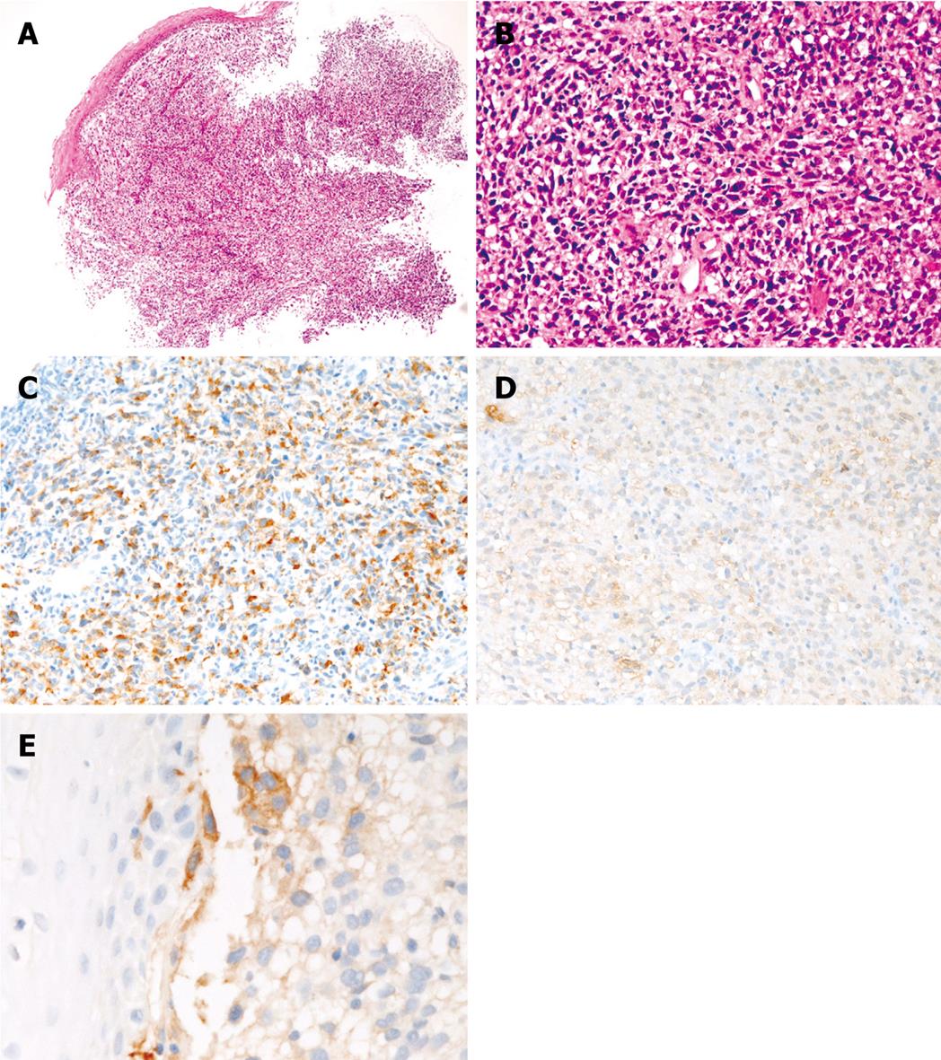
Figure 1 Esophageal amelanotic malignant melanoma in Case 1.
A: Low power view of the biopsy (HE, × 20); B: High power view of the biopsy. Malignant polygonal and spindle cells are seen. No melanin pigment is seen (HE, × 200); C: The tumor cells are positive for melanosome (HMB45). (Immunostaining, × 200); D: The tumor cells are positive for S100 protein (Immunostaining, × 200); E: The tumor cells are focally positive for KIT (Immunostaining, × 200).
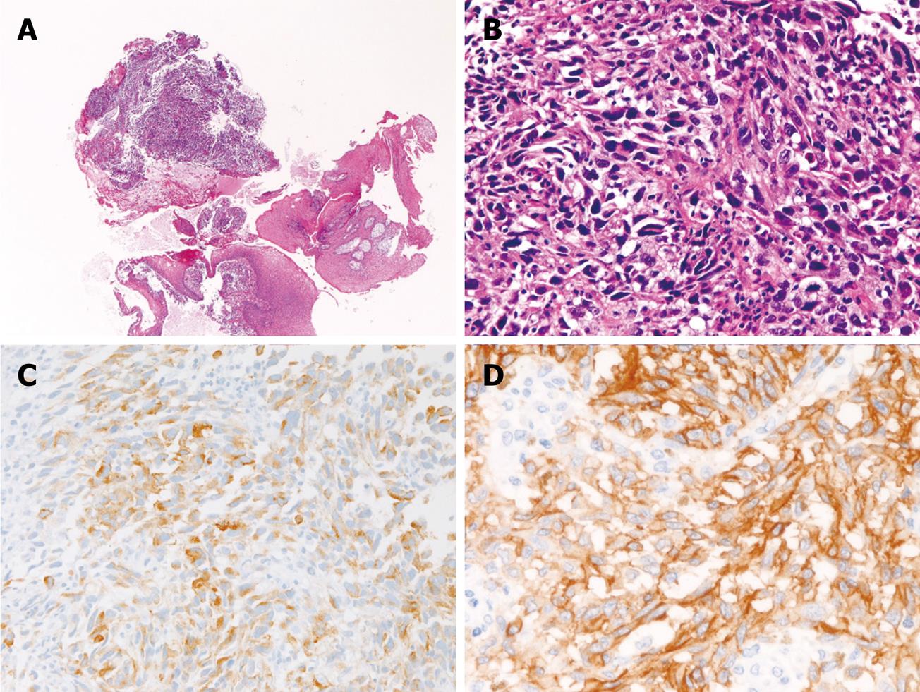
Figure 2 Esophageal amelanotic malignant melanoma in Case 2.
A: Low power view of the biopsy (HE, × 20); B: High power view of the biopsy. Malignant spindle cells are seen. No melanin pigment is seen (HE, × 200); C: The tumor cells are positive for melanosome (HMB45) (Immunostaining, × 200); D: The tumor cells are diffusely positive for KIT (Immunostaining, × 200).
-
Citation: Terada T. Amelanotic malignant melanoma of the esophagus: Report of two cases with immunohistochemical and molecular genetic study of
KIT andPDGFRA . World J Gastroenterol 2009; 15(21): 2679-2683 - URL: https://www.wjgnet.com/1007-9327/full/v15/i21/2679.htm
- DOI: https://dx.doi.org/10.3748/wjg.15.2679


