Copyright
©2009 The WJG Press and Baishideng.
World J Gastroenterol. May 28, 2009; 15(20): 2550-2551
Published online May 28, 2009. doi: 10.3748/wjg.15.2550
Published online May 28, 2009. doi: 10.3748/wjg.15.2550
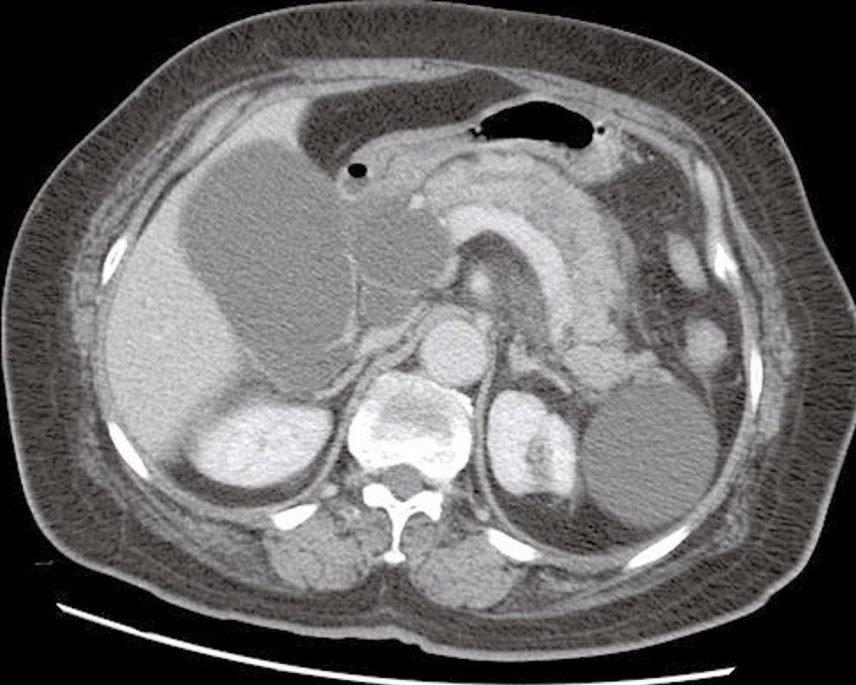
Figure 1 Abdominal CT revealing the dilated common bile duct and pancreatic duct and enlarged pancreatic head.
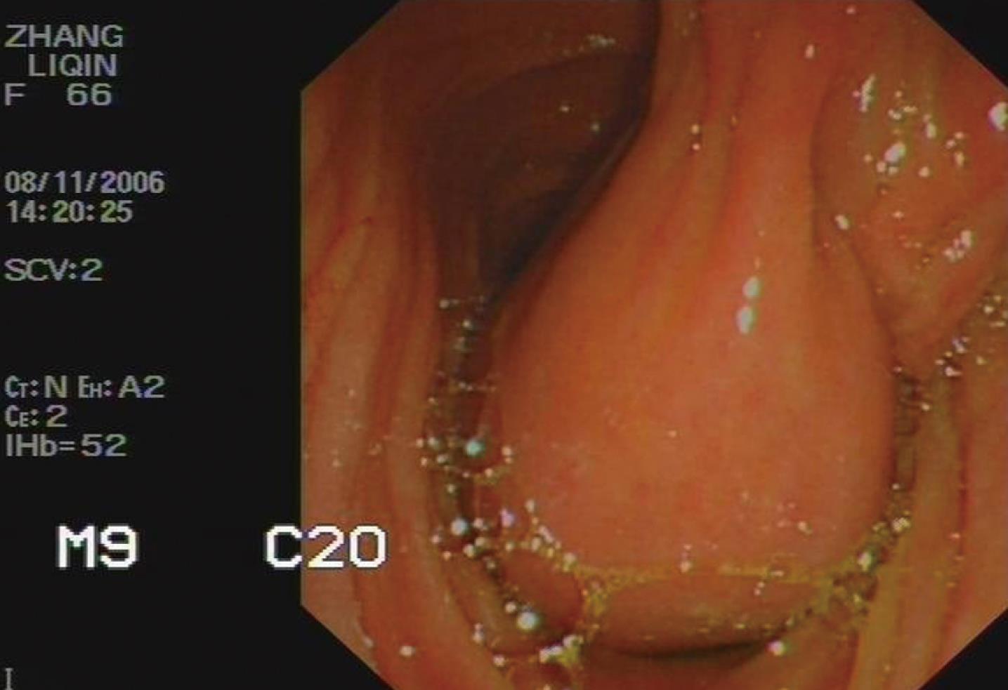
Figure 2 Endoscopy showing a cystic tumor connected to the papilla in duodenum.
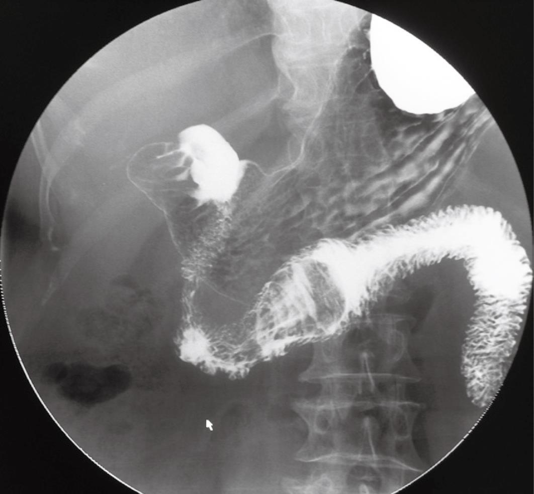
Figure 3 Upper gastrointestinal contrast scan displaying a filling defect in the horizontal part of the duodenum.
- Citation: Wang QG, Zhang ST. A rare case of bile duct cyst. World J Gastroenterol 2009; 15(20): 2550-2551
- URL: https://www.wjgnet.com/1007-9327/full/v15/i20/2550.htm
- DOI: https://dx.doi.org/10.3748/wjg.15.2550


