©2009 The WJG Press and Baishideng.
World J Gastroenterol. Jan 14, 2009; 15(2): 245-247
Published online Jan 14, 2009. doi: 10.3748/wjg.15.245
Published online Jan 14, 2009. doi: 10.3748/wjg.15.245
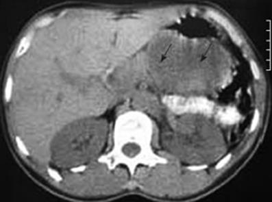
Figure 1 CT scan indicating a tumor at the tail of the pancreas (arrows).
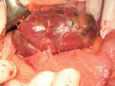
Figure 2 Photograph showing the tumor on the posterior wall of the stomach.
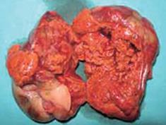
Figure 3 Photograph showing a cross section of the tumor.
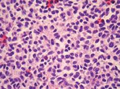
Figure 4 Histological appearance of the diffuse neoplastic infiltration showing rather uniform diffuse small round cells and abortive pseudorosette formation (HE, × 112).
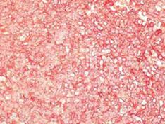
Figure 5 Tumor cells showing strong diffuse membrane immuno-histochemical reactivity with CD99 antibodies [labeled streptavidin biotin (LSAB+) method, 3-amino-9-ethyl carbazole (AEC) visualization, × 112].
- Citation: Colovic RB, Grubor NM, Micev MT, Matic SV, Atkinson HDE, Latincic SM. Perigastric extraskeletal Ewing’s sarcoma: A case report. World J Gastroenterol 2009; 15(2): 245-247
- URL: https://www.wjgnet.com/1007-9327/full/v15/i2/245.htm
- DOI: https://dx.doi.org/10.3748/wjg.15.245


