Copyright
©2009 The WJG Press and Baishideng.
World J Gastroenterol. May 21, 2009; 15(19): 2314-2328
Published online May 21, 2009. doi: 10.3748/wjg.15.2314
Published online May 21, 2009. doi: 10.3748/wjg.15.2314
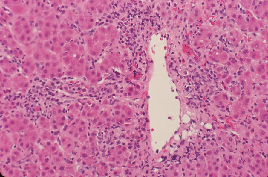
Figure 1 Centrilobular zone 3 necrosis.
Inflammation and hepatocyte drop out are present around a terminal hepatic venule in conjunction with hepatic plate thickening, architectural disorganization, and rosette formation. Centrilobular (perivenular) zone 3 necrosis can be an early acute form of autoimmune hepatitis that can transform to interface hepatitis (HE, × 200).
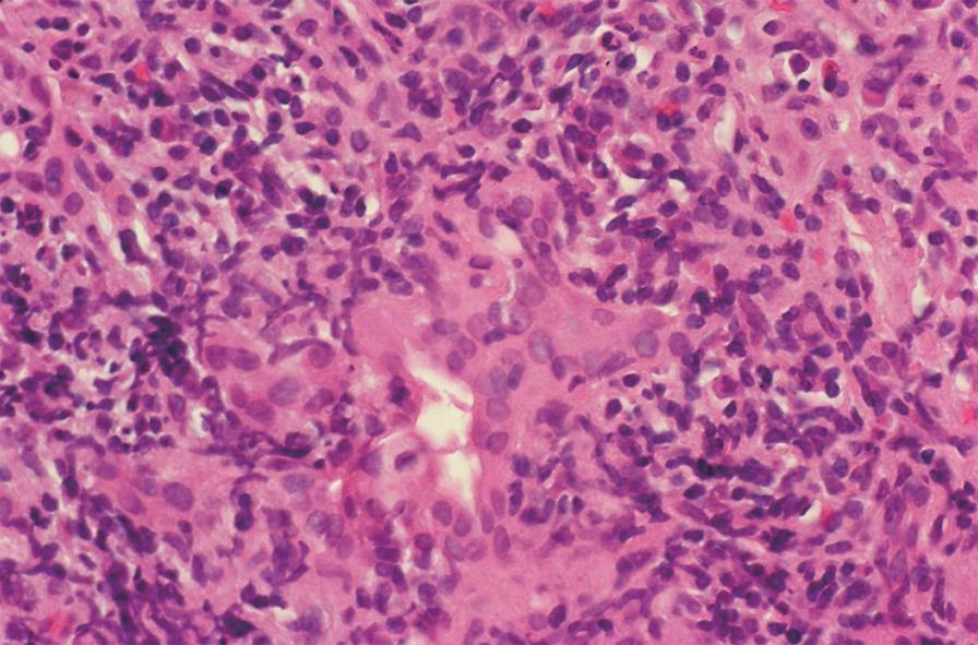
Figure 2 Concurrent pleomorphic cholangitis.
Lymphocytes and histiocytes surround, infiltrate and damage an interlobular bile duct. Bile duct injury in the absence of cholestatic clinical and laboratory manifestations may represent collateral injury that is transient (HE, × 400).
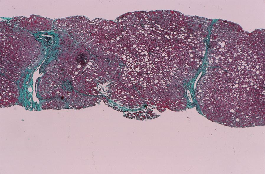
Figure 3 Steatosis.
Macrovesicular steatosis is the predominant histological feature after corticosteroid treatment. Fatty changes may be present before or during corticosteroid treatment and perpetuate or extend the laboratory indices of liver inflammation (Trichrome stain, × 40).
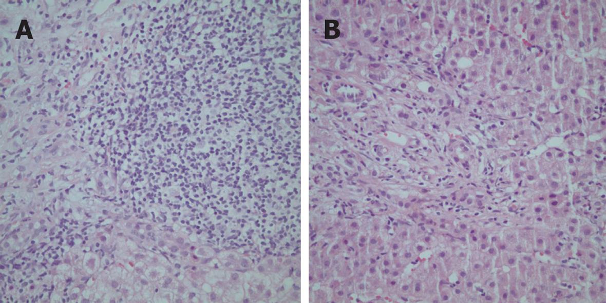
Figure 4 Histological features of a Turkish patient with the “overlap syndrome” (autoimmune hepatitis and primary biliary cirrhosis) characterized by heavy portal infiltration with lymphocytes and plasma cells (A) and bile ductular proliferation and ductopenia (B) (HE, × 200).
- Citation: Czaja AJ, Bayraktar Y. Non-classical phenotypes of autoimmune hepatitis and advances in diagnosis and treatment. World J Gastroenterol 2009; 15(19): 2314-2328
- URL: https://www.wjgnet.com/1007-9327/full/v15/i19/2314.htm
- DOI: https://dx.doi.org/10.3748/wjg.15.2314


