©2009 The WJG Press and Baishideng.
World J Gastroenterol. May 7, 2009; 15(17): 2109-2115
Published online May 7, 2009. doi: 10.3748/wjg.15.2109
Published online May 7, 2009. doi: 10.3748/wjg.15.2109
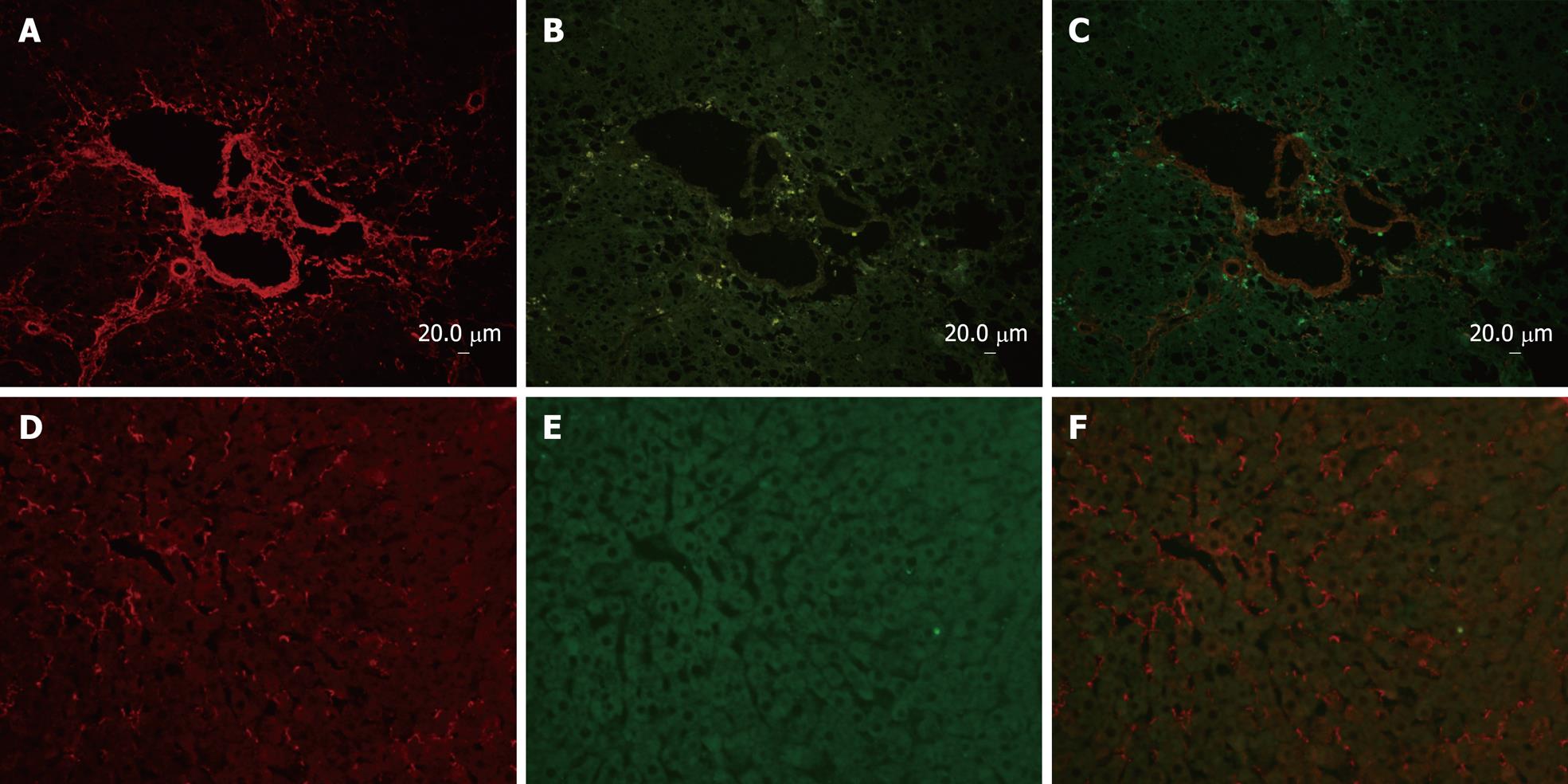
Figure 1 Co-immunofluoresence of p90RSK and collagen type I in fibrotic liver and normal liver.
Sections (A-C) are from rat liver with intraperitoneal injection of DMN for 3 wk, and sections (D-F) are from normal livers as control. Sections of fibrotic liver mostly demonstrate that collagen type I (rhodamine) and p90RSK (FITC) immunoreactivity were both present around central veins as well as in the interstitium, and up-regulated in fibrotic liver. Sections of normal liver mostly demonstrate that collagen type I (rhodamine) immunoreactivity was less in normal liver than in fibrotic liver and p90RSK (FITC) was hardly observed in nomal liver simultaneously.
Figure 2 Cellular localization of p90RSK in fibrotic liver and co-immunofluoresence and confocal microscopy of p90RSK and αSMA in fibrotic liver.
A: α-SMA (rhodamine)-positive cell represent the activated HSC, which deposited in interstitium; B: P90RSK (FITC)-positive cell were also present in interstitium; C: The yellow areas on the merged image show co-localization of α-SMA and p90RSK.
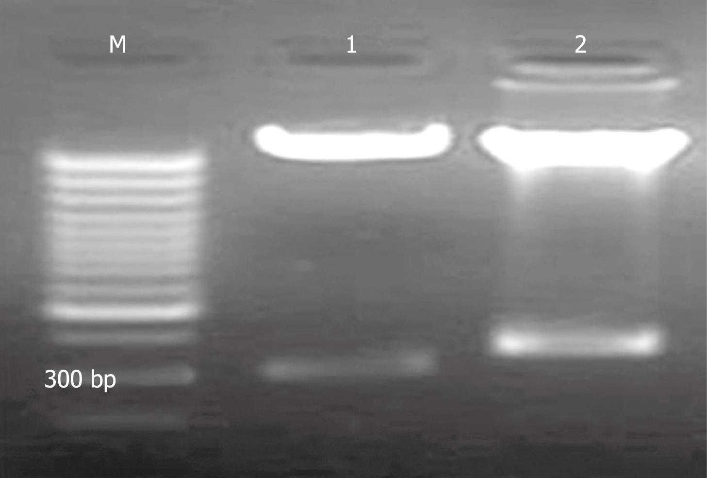
Figure 3 Agarose gel electrophoresis of restriction enzyme digestion of pAV-siRSK.
M: 1 kb marker; 1: Restriction enzyme digestion product of pAVU6+27 was about 300 bp; 2: Restriction enzyme digestion product of pAV-siRSK. The siRNA of p90RSK was designed of 52 bp.
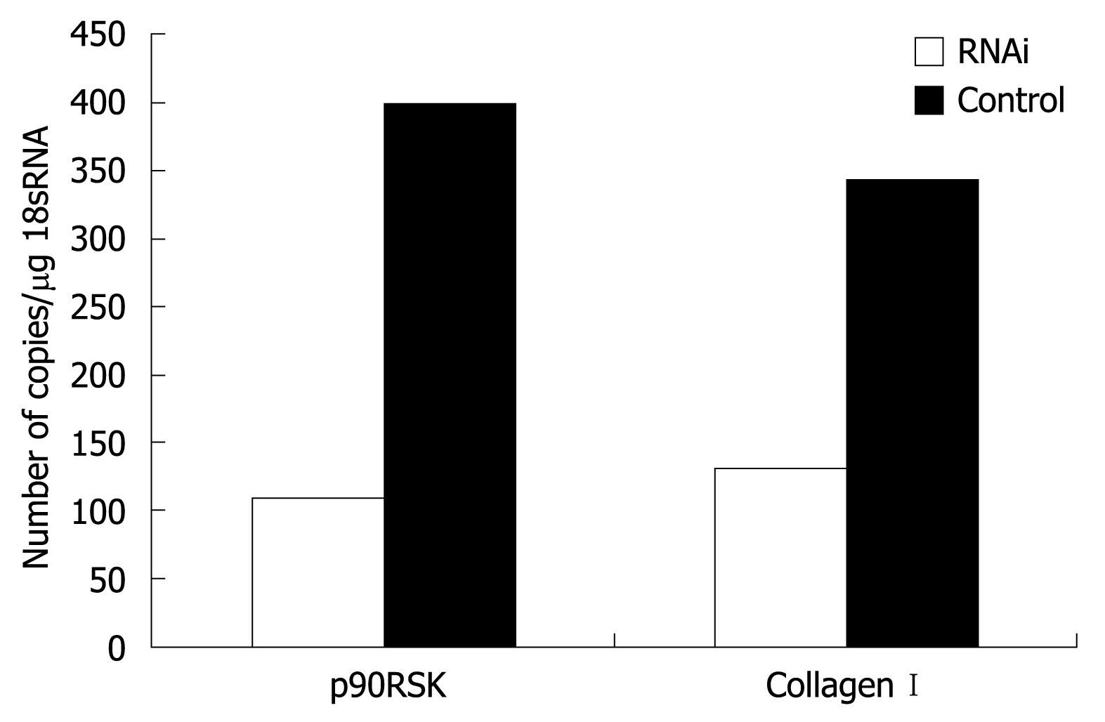
Figure 4 RT-PCR assessment of p90RSK and collagen type I in HSC-T6 transfected with or without pAVU6-siRSK.
Quantification of p90RSK and collagen type I normalized to 18S RNA in HSC-T6 cells transfected with pAVU6-siRSK decreased 72.6% and 61.8%, respectively, compared with control (P < 0.01). Results are expressed as mean ± SE of three separate experiments.
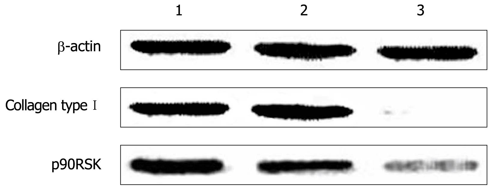
Figure 5 Western blotting analysis of p90RSK, collagen type I in HSC-T6 cells transfected with or without pAVU6-siRSK.
β-actin provided as an inner control. 1: Normal HSC-T6 cells; 2: HSC-T6 transfected with empty plasmid; 3: HSC-T6 transfected with pAVU6-siRSK. With p90RSK siRNA, the expression of p90RSK decreased 75.6% compared with controls, and the expression of collagen type I decreased 89.1% accordingly (P < 0.01). Results are expressed as mean ± SE of three separate experiments.
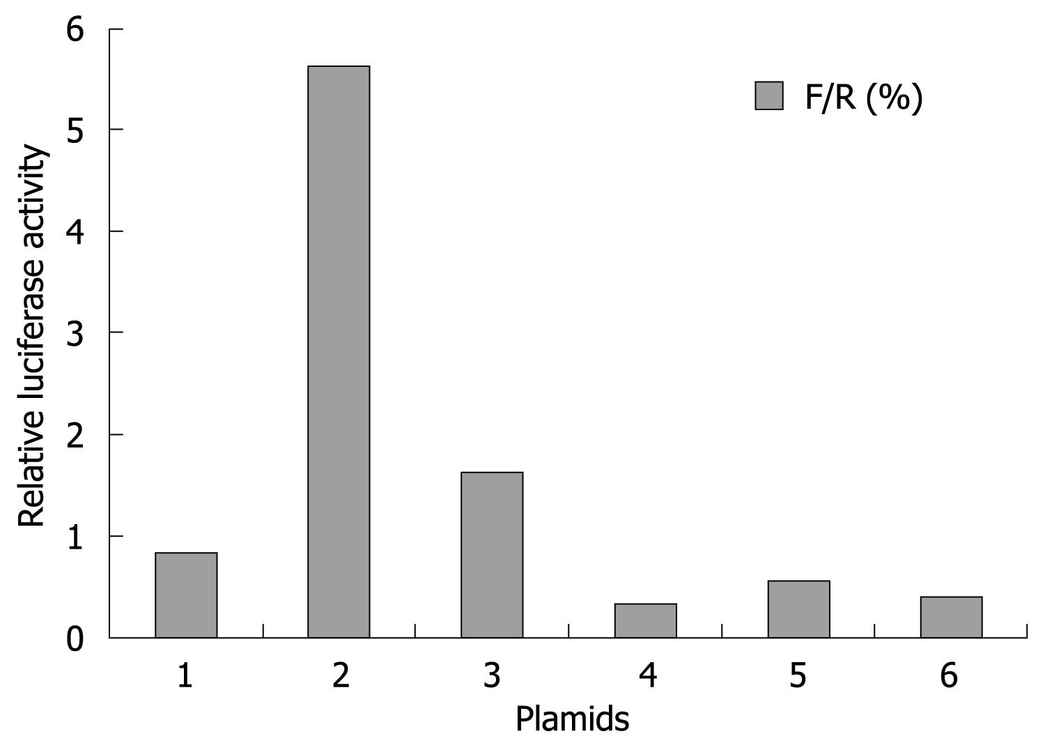
Figure 6 Effect of p90RSK on collagen type I promoter activity.
HSCs transfected with the pAVU6-siRSK (bar 3, to decrease p90RSK expression), pMT2·RSK1 (bar 5, to increase p90RSK expression) showed no alteration of collagen type I promoter activity compared to control cells sham-transfected with pAVU6+27 (bar 4), pMT2 (bar 6), respectively. HSCs transfected with collagen type I luciferase reporter construct (bar 1) as normal control and Renilla luciferase expression construct (bar 2) as positive control. After 24 h incubation, the luciferase activity was determined. Data represent luciferase activity relative to the control (bar 1) and are expressed as mean ± SD of three separate experiments.
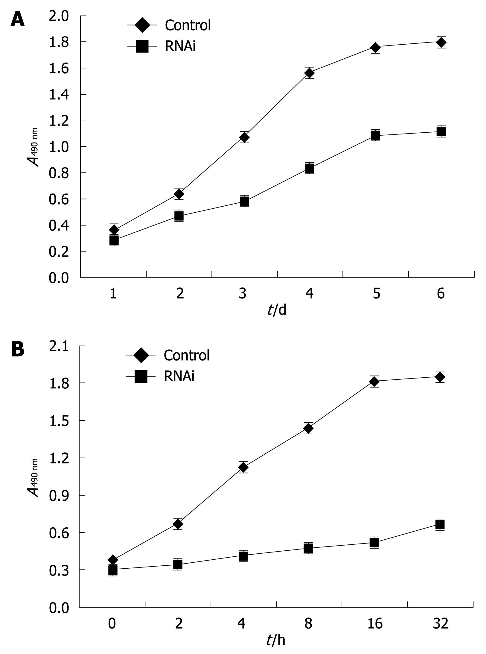
Figure 7 Effect of p90RSK siRNA on the proliferation of HSCs.
A: p90RSK siRNA inhibited the proliferation of HSC-T6 cells. Cell growth curves of the recombinant cells with or without p90RSK siRNA were analyzed by MTS conversion; B: p90RSK siRNA abolished the effect of PDGF-BB on the proliferation of HSCs. rhPDGF-BB was added to the medium at a final concentration of 20 &mgr;g/L in HSC-T6 cells transfected with pAVU6-siRSK (RNAi), which showed decreased proliferative activity compared to sham transfected control cells (control). Each sample was tested in triplicate and error bars were included.
- Citation: Yang MF, Xie J, Gu XY, Zhang XH, Davey AK, Zhang SJ, Wang JP, Zhu RM. Involvement of 90-kuD ribosomal S6 kinase in collagen type I expression in rat hepatic fibrosis. World J Gastroenterol 2009; 15(17): 2109-2115
- URL: https://www.wjgnet.com/1007-9327/full/v15/i17/2109.htm
- DOI: https://dx.doi.org/10.3748/wjg.15.2109


