©2009 The WJG Press and Baishideng.
World J Gastroenterol. Apr 7, 2009; 15(13): 1636-1640
Published online Apr 7, 2009. doi: 10.3748/wjg.15.1636
Published online Apr 7, 2009. doi: 10.3748/wjg.15.1636
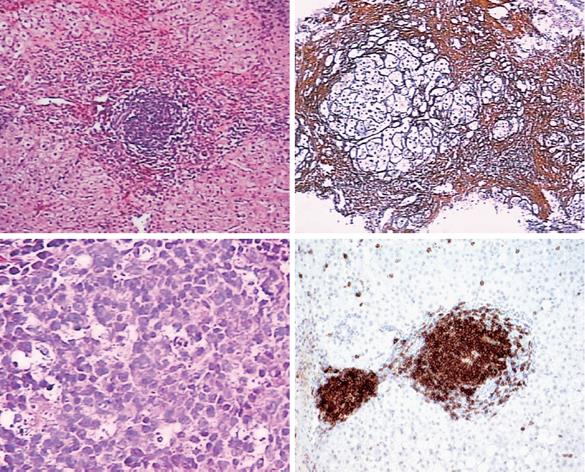
Figure 1 Histological appearances of a B-cell lymphoma, best classified as a marginal zone lymphoma involving the liver.
A, B: Low power view of liver core biopsy shows chronic hepatitis and marked portal lymphoid infiltrates. C: High power view of liver core biopsy shows a monotonous portal lymphoid infiltrate composed of small lymphocytes with moderate amount of clear cytoplasm (so-called monocytoid appearance). Note that the infiltrate does not involve the biliary epithelium. D: CD20 immunostain shows that virtually all of the lymphoid cells are B-cells.
- Citation: Fan HB, Zhu YF, Chen AS, Zhou MX, Yan FM, Ma XJ, Zhou H. B-cell clonality in the liver of hepatitis C virus-infected patients. World J Gastroenterol 2009; 15(13): 1636-1640
- URL: https://www.wjgnet.com/1007-9327/full/v15/i13/1636.htm
- DOI: https://dx.doi.org/10.3748/wjg.15.1636


