©2009 The WJG Press and Baishideng.
World J Gastroenterol. Apr 7, 2009; 15(13): 1587-1593
Published online Apr 7, 2009. doi: 10.3748/wjg.15.1587
Published online Apr 7, 2009. doi: 10.3748/wjg.15.1587
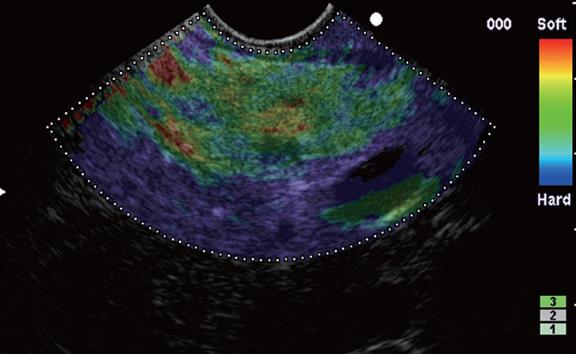
Figure 1 Elastographic image showing homogenous soft tissue corresponding to normal tissue.
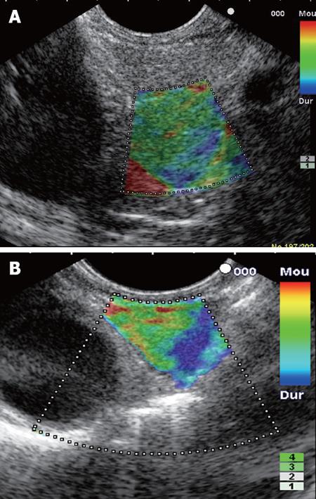
Figure 2 Elastographic image.
A: Heterogenous soft tissue corresponding to fibrosis (benign nodule in patient who had an acute pancreatitis 2 mo before); B: Heterogenous soft tissue corresponding to inflammatory tissue (benign lymph node).
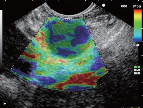
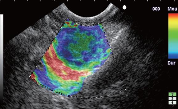
Figure 3 Elastographic image showing mixed hard and soft tissue (“honeycombed pattern”) making the interpretation difficult.
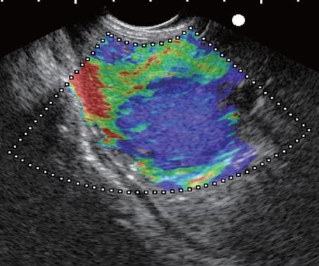
Figure 4 Elastographic image showing mainly hard tissue with a small soft central area corresponding to a malignant hypervascularized lesion (pancreatic neuroendocrine tumor).
Figure 5 Elastographic image showing mainly hard tissue with areas of heterogenous soft tissue corresponding to an advanced malignant lesion with necrotic areas (pancreatic adenocarcinoma).
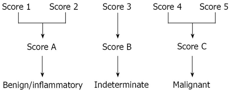
Figure 6 Elastography score.
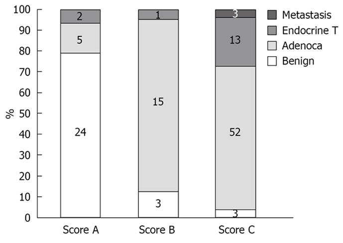
Figure 7 Pancreatic masses: elastography score and final histology.
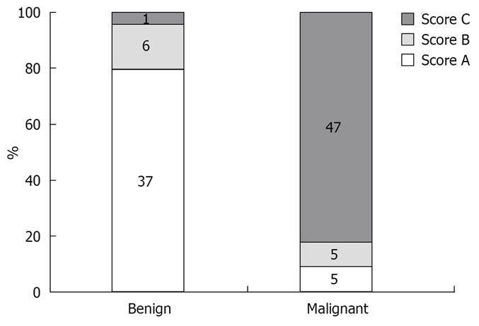
Figure 8 Lymph nodes: elastography score and final histology.
- Citation: Giovannini M, Botelberge T, Bories E, Pesenti C, Caillol F, Esterni B, Monges G, Arcidiacono P, Deprez P, Yeung R, Schimdt W, Schrader H, Szymanski C, Dietrich C, Eisendrath P, Van Laethem JL, Devière J, Vilmann P, Saftoiu A. Endoscopic ultrasound elastography for evaluation of lymph nodes and pancreatic masses: A multicenter study. World J Gastroenterol 2009; 15(13): 1587-1593
- URL: https://www.wjgnet.com/1007-9327/full/v15/i13/1587.htm
- DOI: https://dx.doi.org/10.3748/wjg.15.1587


