Copyright
©2009 The WJG Press and Baishideng.
World J Gastroenterol. Mar 14, 2009; 15(10): 1273-1275
Published online Mar 14, 2009. doi: 10.3748/wjg.15.1273
Published online Mar 14, 2009. doi: 10.3748/wjg.15.1273
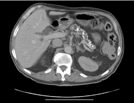
Figure 1 Abdominal computed tomography image showing gross pancreatic duct dilatation and extensive pancreatic calcification as well as bile duct dilatation.
No distinct mass was seen.
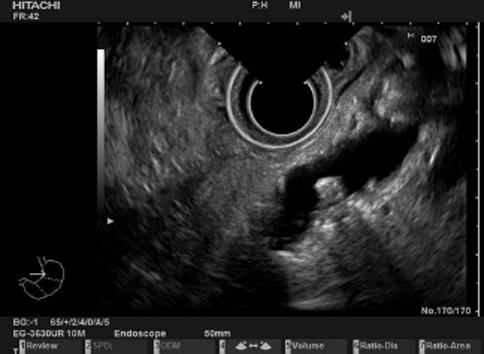
Figure 2 Endoscopic ultrasound revealing a dilated pancreatic duct (> 10 mm in diameter) with mural irregularities, calculi within the pancreatic duct, and an atrophic pancreas.
- Citation: Kalaitzakis E, Braden B, Trivedi P, Sharifi Y, Chapman R. Intraductal papillary mucinous neoplasm in chronic calcifying pancreatitis: Egg or hen? World J Gastroenterol 2009; 15(10): 1273-1275
- URL: https://www.wjgnet.com/1007-9327/full/v15/i10/1273.htm
- DOI: https://dx.doi.org/10.3748/wjg.15.1273


