©2008 The WJG Press and Baishideng.
World J Gastroenterol. Feb 21, 2008; 14(7): 1038-1043
Published online Feb 21, 2008. doi: 10.3748/wjg.14.1038
Published online Feb 21, 2008. doi: 10.3748/wjg.14.1038
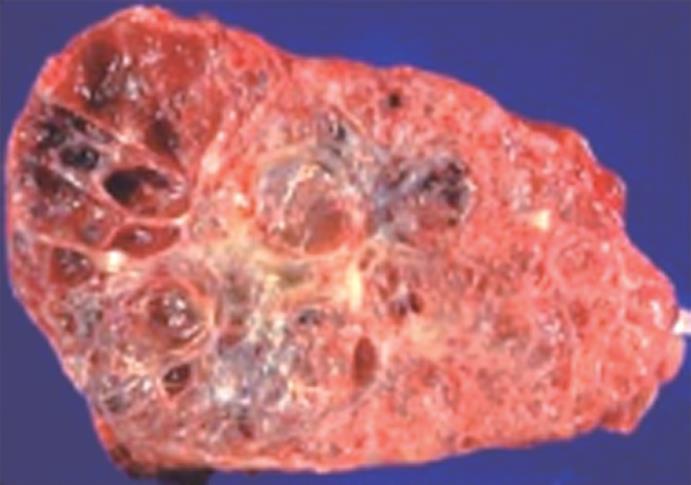
Figure 1 Gross surgical pathology specimen of a serous cystadenoma.
Note the presence of a honeycomb appearance of the lesion.
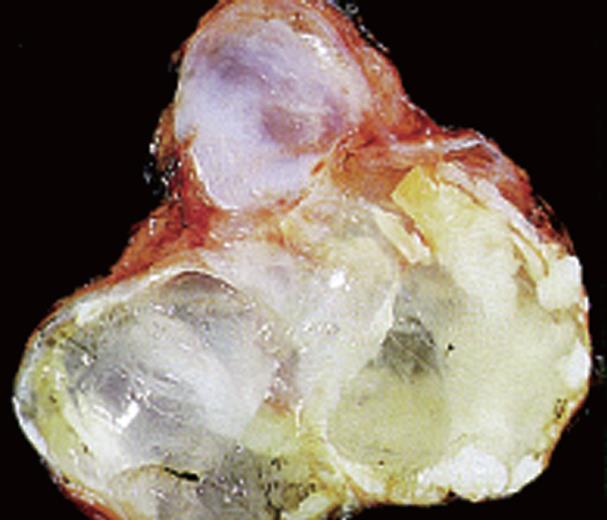
Figure 2 Gross surgical pathology specimen of a mucinous cystadenoma.
The cyst cavities are filled with a clear viscous fluid.
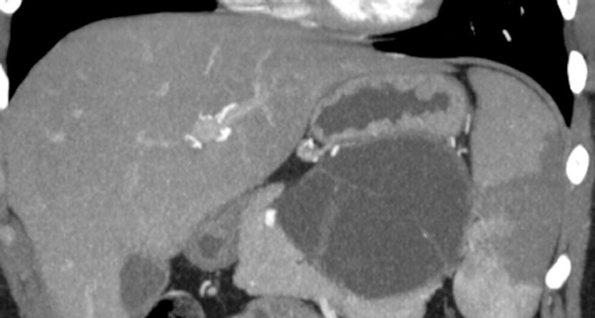
Figure 3 Reformatted CT demonstrating a mucinous cystic neoplasm indenting the stomach.
Note the presence of septations.
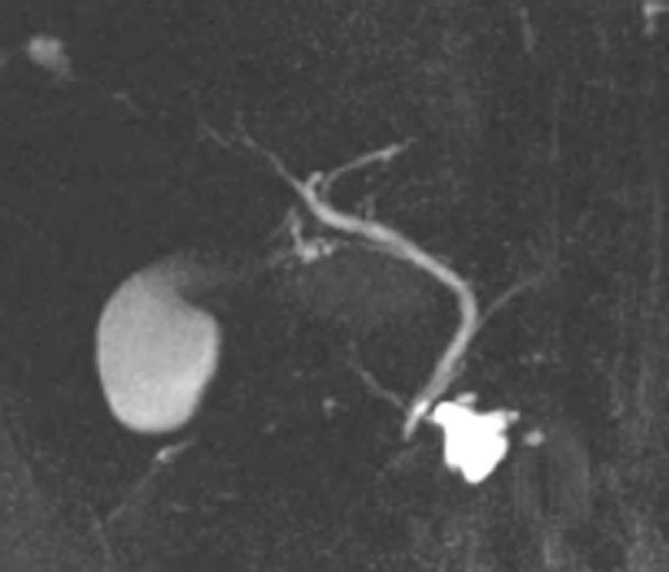
Figure 4 MRCP of side branch IPMN.
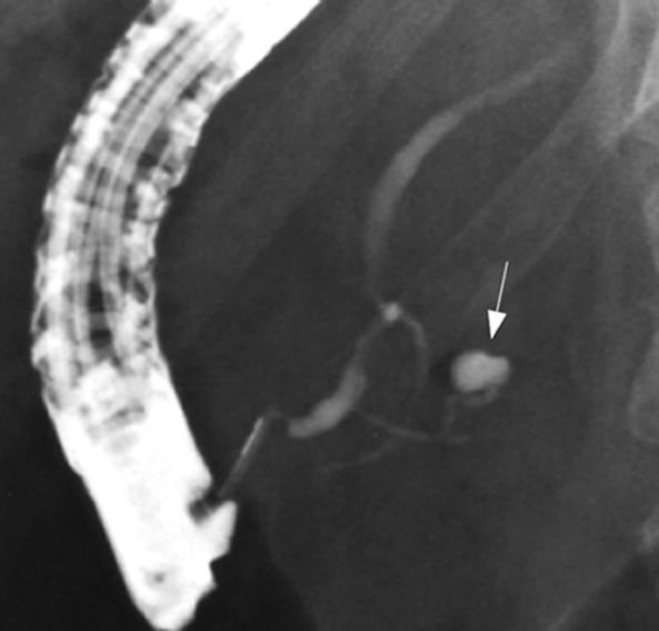
Figure 5 Pancreatogram during ERCP demonstrating a small side branch IPMN.
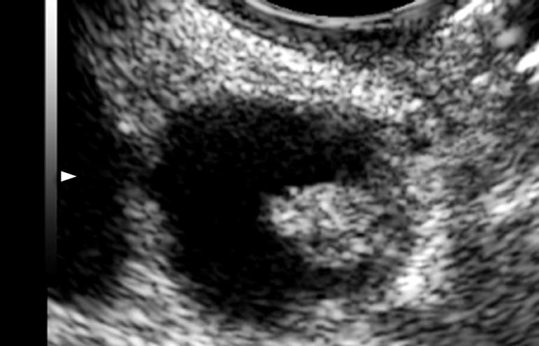
Figure 6 Linear EUS Imaging of a mucinous cystic neoplasm containing a mural nodule.
- Citation: Brugge WR. Diagnosis and management of relapsing pancreatitis associated with cystic neoplasms of the pancreas. World J Gastroenterol 2008; 14(7): 1038-1043
- URL: https://www.wjgnet.com/1007-9327/full/v14/i7/1038.htm
- DOI: https://dx.doi.org/10.3748/wjg.14.1038


