Copyright
©2008 The WJG Press and Baishideng.
World J Gastroenterol. Feb 21, 2008; 14(7): 1016-1022
Published online Feb 21, 2008. doi: 10.3748/wjg.14.1016
Published online Feb 21, 2008. doi: 10.3748/wjg.14.1016
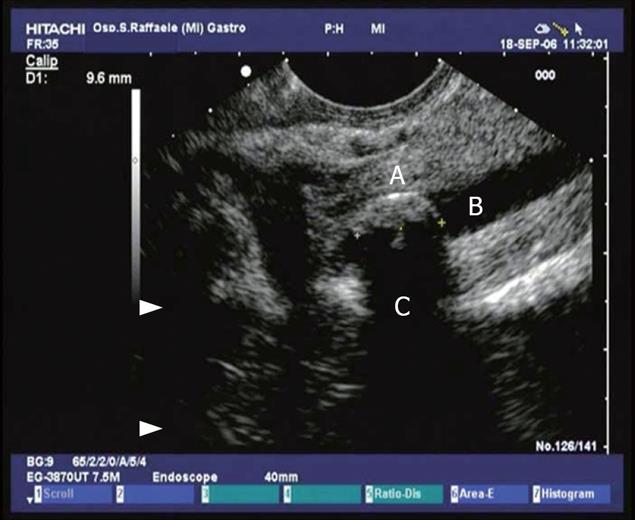
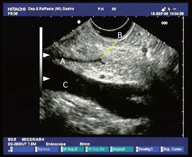
Figure 1 Conventional endosonographic imaging (7.
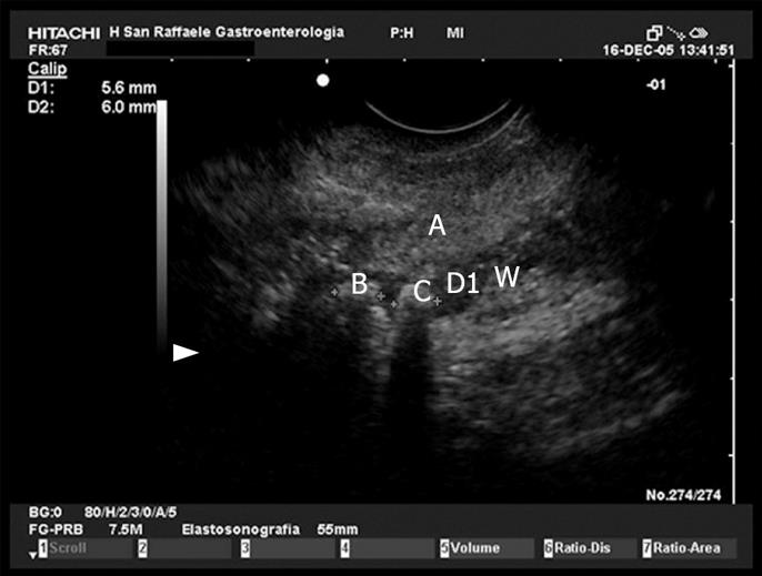
5 MHz) of choledocholithiasis. A 9.6 mm stone (A) is seen as an hyperechoic structure with acoustic shadowing (C) in the intrapancreatic tract of the common bile duct (B).
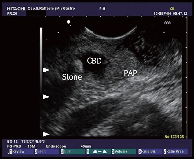
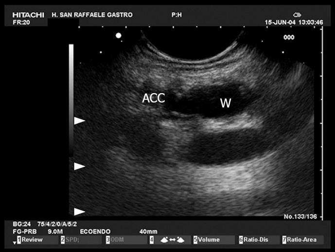
Figure 2 Common bile duct (CBD) stone with dilation of the biliary tract.
PAP: Vater papilla.
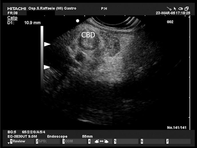
Figure 3 View of the proximal tract of the common bile duct (CBD) from the duodenum: the duct is dilated and completely obstructed by minuscule concrements (microlithiasis).
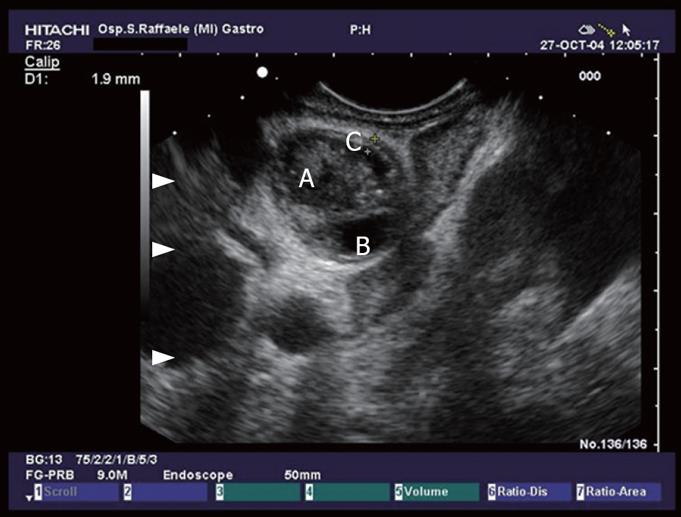
Figure 4 Hepatic ilum seen from the duodenal bulb: common bile duct with sludge (A), thickening of the CBD wall (C), and cystic duct (B).
Figure 5 Pancreas divisum.
The pancreatic duct at the istmus moves towards the minor papilla (B), (A) dorsal pacreatic duct, (C) superior mesenteric vein.
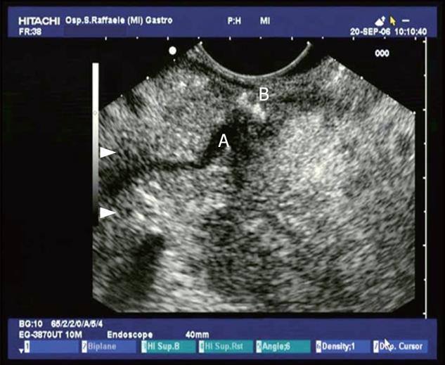
Figure 6 A view of Santorini’s duct (A) from the duodenal bulb, with millimetres stones in the retropapillary region (B) (minor papilla).
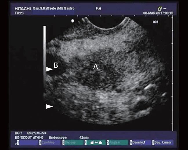
Figure 7 A malignant lesion (A) in the pancreatic head occluding the main pancreatic duct wich is dilated (B).
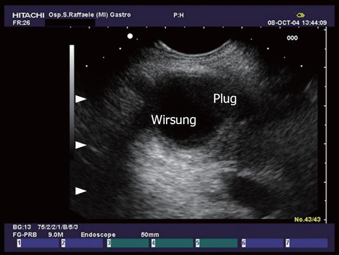
Figure 8 Dilation of the main pancreatic duct and collateral duct with evidence of plugs in the lumen in a patient with IPMT.
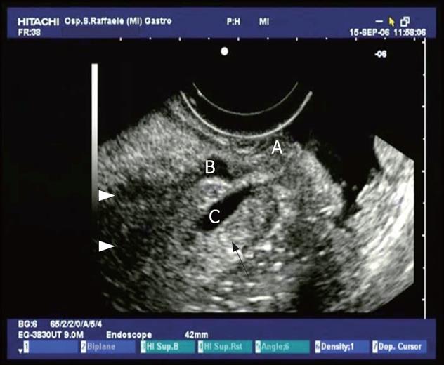
Figure 9 IPMT.
A small cystic lesion (C) in retropapillary region (A), that seems to communicate with main pancreatic duct (B).
Figure 10 IPMT.
Dilation of the main pancreatic duct and accessory duct. The sorrounding parenchyma is hypotrofic, hypoechoic.
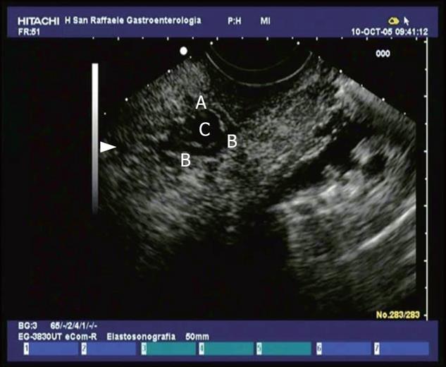
Figure 11 Normal ampulla.
The ampulla (A) is seen as an hypoechoic structure from which the two ducts, the common bile duct (B) and the Wirsung (C) can be followed into the pancreatic head.
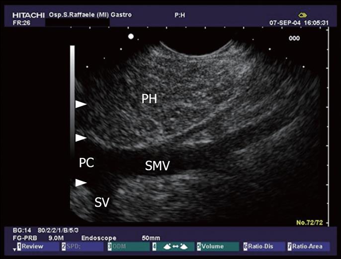
Figure 12 The pancreatic head (PH) seen from the duodenum.
Minimal changes like echogenic strains in the echo texture of the pancreatic parenchyma. Portal confluence (PC) with its vessels: the superior mesenteric vein (SMV) and splenic vein (SV).
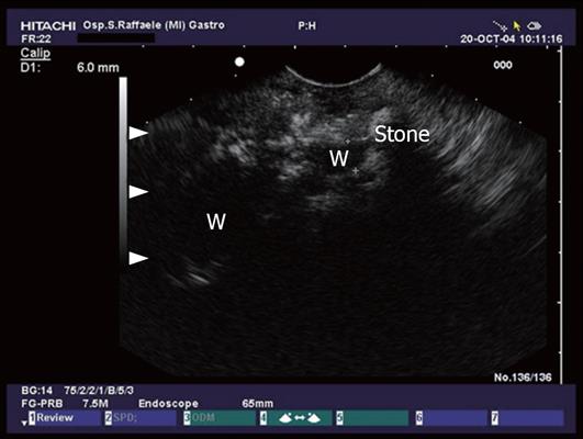
Figure 13 Advanced chronic pancreatitis with calcifications, pancreatic stone and dilation of the pancreatic duct.
Figure 14 Pancreatic head (A) and pancreatic duct stones (B and C).
- Citation: Petrone MC, Arcidiacono PG, Testoni PA. Endoscopic ultrasonography for evaluating patients with recurrent pancreatitis. World J Gastroenterol 2008; 14(7): 1016-1022
- URL: https://www.wjgnet.com/1007-9327/full/v14/i7/1016.htm
- DOI: https://dx.doi.org/10.3748/wjg.14.1016


