©2008 The WJG Press and Baishideng.
World J Gastroenterol. Feb 14, 2008; 14(6): 876-883
Published online Feb 14, 2008. doi: 10.3748/wjg.14.876
Published online Feb 14, 2008. doi: 10.3748/wjg.14.876
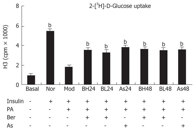
Figure 1 Insulin-induced 2-deoxy-[3H]-D-Glucose uptake in 3T3-L1 adipocytes .
3T3-L1 preadipocytes (5 × 10⁵/well) were differentiated to adipocytes in a 24-well plate. After serum-starvation in 0.2% BSA DMEM overnight, the cells were incubated in 0.2% BSA DMEM containing 0.5 mmol/L PA or (and) 1 &mgr;mol/L, 10 &mgr;mol/L Ber or 5 mmol/L Aspirin for 24 h or 48 h. Then, the cells were incubated in 1 mL KRP-HEPES without or with 100 nmol/L insulin for 30 min at 37°C after washed three times in KRP-HEPES buffer. Next, the cells were incubated in 1 mL KRP-HEPES containing 0.5 &mgr;Ci/mL 2-deoxy-D-[3H] glucose for 10 min at 37°C. Finally, the cells were washed three times in ice-cold PBS and solubilized in 1ml of 0.1 mol/L NaOH for 2 h. Radioactivity was determined by liquid scintillation spectrometry. Nonspecific deoxyglucose uptake was measured in the presence of 20 &mgr;mol/L cytochalasin B and specific glucose uptake was detected from the subtracted total uptake. Three replicate wells were set up and each experiment was repeated three times. bP < 0.01, vs Mod group.
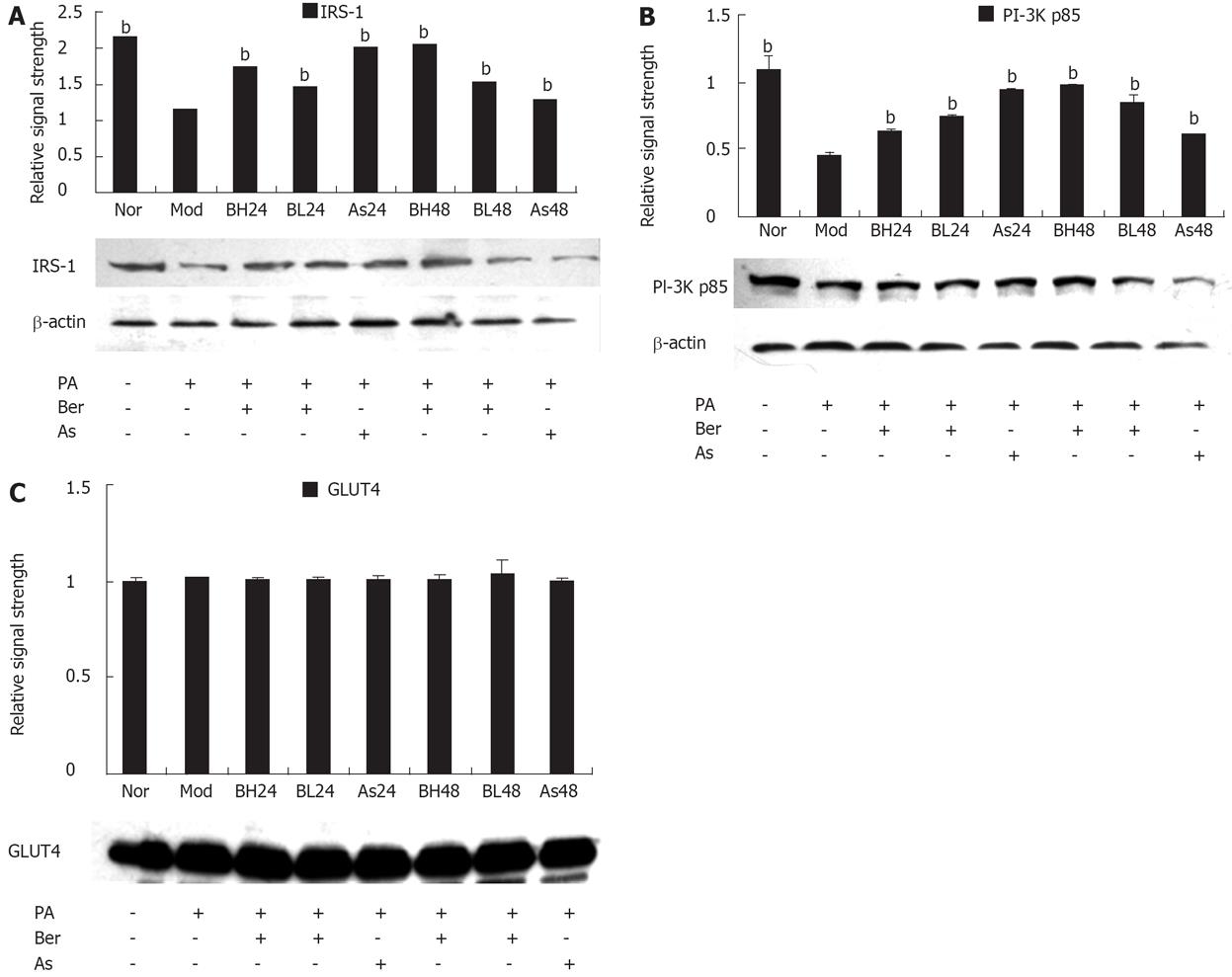
Figure 2 A: Effect of Ber on the IRS-1 Protein Expression in 3T3-L1 adipocytes.
Fully differentiated cells were cultured for 12 h in serum-free DMEM with 0.2% BSA and then they were, respectively, cultured for 24 h in DMEM containing 0.5 mmol/L PA and 1% BSA (Mod); or cultured for 24 and 48 h in DMEM containing 0.5 mmol/L PA, 10 &mgr;mol/L Ber, 1% BSA (BH24, BH48); or in DMEM containing 0.5 mmol/L PA, 1 &mgr;mol/L Ber, 1% BSA for 24 h and 48 h (BL24, BL48); or cultured in DMEM containing 0.5 mmol/L PA, 5 mmol/L aspirin, 1% BSA for 24 h and 48 h (As24, As48); or in DMEM containing 1% BSA for 24 h (Nor). IRS-1 proteins were determined in the whole cell lysate by immunoblotting. Each experiment was repeated three times. bP < 0.01 vs Mod group; B: Effect of Ber on the PI-3K p85 Protein Expression in 3T3-L1 adipocytes. Fully differentiated cells were cultured for 12 h in serum-free DMEM with 0. 2% BSA and then they were, respectively, cultured for 24 h in DMEM containing 0.5 mmol/L PA and 1% BSA (Mod); or cultured for 24 and 48 h in DMEM containing 0.5 mmol/L PA, 10 &mgr;mol/ L Ber, 1% BSA (BH24, BH48); or in DMEM containing 0.5 mmol/L PA,1 &mgr;mol/ L Ber, 1% BSA for 24 h and 48 h (BL24, BL48); or cultured in DMEM containing 0.5 mmol/L PA, 5 mmol/L aspirin, 1% BSA for 24 h and 48 h (As24, As48); or in DMEM containing 1% BSA for 24 h (Nor). Insulin was used at 100 nmol/L for 30 min. PI-3K p85 proteins were determined in the whole cell lysate by immunoblotting. Each experiment was repeated three times. bP < 0.01 vs Mod group; C: Effect of Ber on the GLUT4 Protein Expression in 3T3-L1 Adipocytes. Fully differentiated cells were cultured for 12 h in serum-free DMEM with 0.2% BSA and then they were, respectively, cultured for 24 h in DMEM containing 0.5 mmol/L PA and 1% BSA (Mod); or cultured for 24 and 48 h in DMEM containing 0.5 mmol/L PA, 10 &mgr;mol/L Ber, 1% BSA (BH24, BH48); or in DMEM containing 0.5 mmol/L PA, 1 &mgr;mol/L Ber, 1% BSA for 24 h and 48 h (BL24, BL48); or cultured in DMEM containing 0.5 mmol/L PA, 5 mmol/L aspirin, 1% BSA for 24 h and 48 h (As24, As48); or in DMEM containing 1% BSA for 24 h (Nor). GLUT4 proteins were determined in the whole cell lysate by immunoblotting. Each experiment was repeated three times.
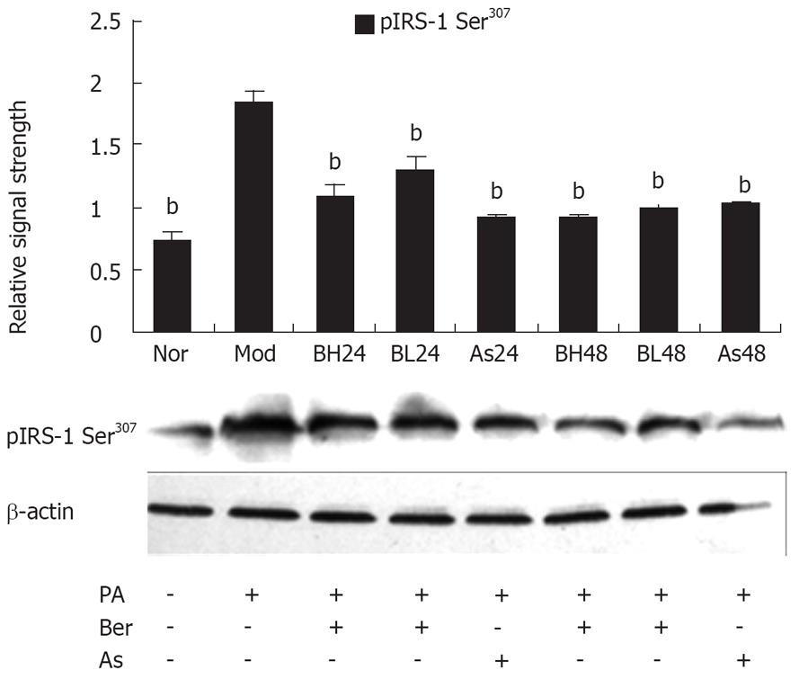
Figure 3 Effect of Ber on the IRS-1 Ser307 Phosphorylation in 3T3-L1 Adipocytes.
Fully differentiated cells were cultured for 12 h in serum-free DMEM with 0.2% BSA and then they were, respectively, cultured for 24 h in DMEM containing 0.5 mmol/L PA and 1% BSA (Mod); or cultured for 24 and 48 h in DMEM containing 0.5 mmol/L PA, 10 &mgr;mol/ L Ber, (1% BSA, BH24) BH48; or in DMEM containing 0.5 mmol/L PA, 1 &mgr;mol/ L Ber, 1% BSA for 24 h and 48 h (BL24, BL48); or cultured in DMEM containing 0.5 mmol/L PA, 5 mmol/L aspirin, 1% BSA for 24 h and 48 h (As24, As48); or in DMEM containing 1% BSA for 24 h (Nor). IRS-1 phosphorylation was determined in the whole cell lysate by immunoblotting with the phosopho-spcific IRS-1(Ser307) antibody. Each experiment was repeated three times. bP < 0.01, vs Mod group.
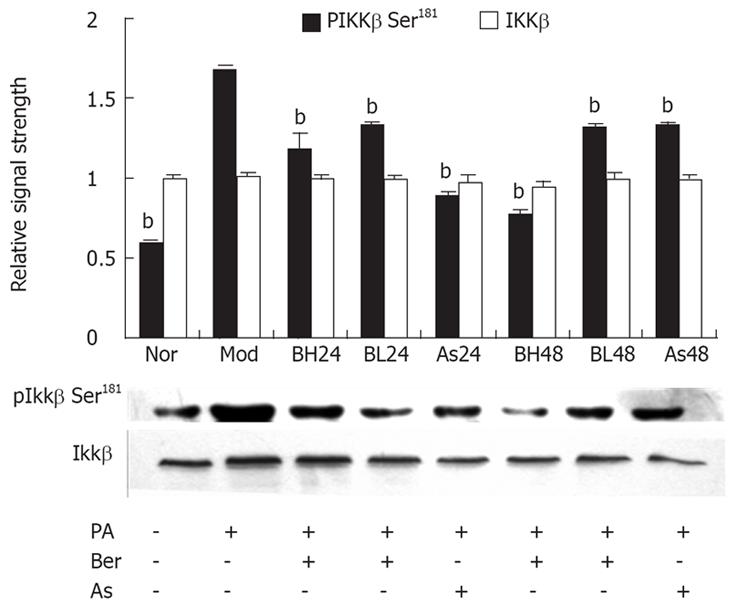
Figure 4 Effect of Ber on the IKKβ Ser181 Phosphorylation and IKKβ protein expression in 3T3-L1 Adipocytes.
Fully differentiated cells were cultured for 12 h in serum-free DMEM with 0.2% BSA and then they were, respectively, cultured for 24 h in DMEM containing 0.5 mmol/L PA and 1% BSA (Mod); or cultured for 24 and 48 h in DMEM containing 0.5 mmol/L PA, 10 &mgr;mol/L Ber, 1% BSA, BH24, BH48; or in DMEM containing 0.5 mmol/L PA, 1 &mgr;mol/L Ber, 1% BSA for 24 h and 48 h (BL24, BL48); or cultured in DMEM containing 0.5 mmol/L PA, 5 mmol/L aspirin, 1% BSA for 24 h and 48 h (As24, As48); or in DMEM containing 1% BSA for 24 h (Nor). IKKβ phosphorylation and IKKβ protein expression were determined in the whole cell lysate by immunoblotting with the phosopho-spcific IKKβ (Ser181) antibody and IKKβ antibody respectively. Each experiment was repeated three times. bP < 0.01, as compared with Mod group.
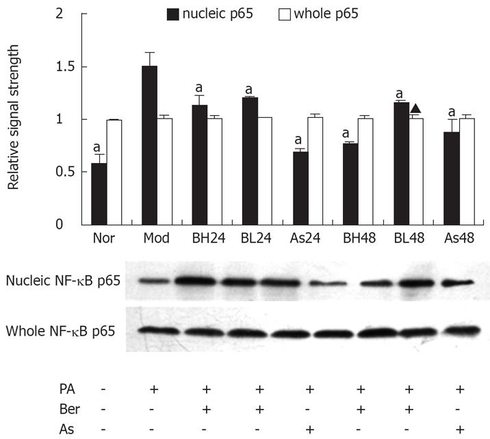
Figure 5 Effect of Ber on the whole NF-κB p65 and nucleic NF-κB p65 protein expression in 3T3-L1 Adipocytes.
Fully differentiated cells were cultured for 12 h in serum-free DMEM with 0.2% BSA and then they were, respectively, cultured for 24 h in DMEM containing 0.5 mmol/L PA and 1% BSA (Mod); or cultured for 24 and 48 h in DMEM containing 0.5 mmol/L PA, 10 &mgr;mol/L Ber,1% BSA (BH24, BH48); or in DMEM containing 0.5 mmol/L PA, 1 &mgr;mol/L Ber, 1% BSA for 24 h and 48 h (BL24, BL48); or cultured in DMEM containing 0.5 mmol/L PA, 5 mmol/L aspirin, 1% BSA for 24 h and 48 h (As24, As48); or in DMEM containing 1% BSA for 24 h (Nor).whole NF-κB p65 and nucleic NF-κB p65 protein expression were determined in the whole cell lysate and nucleic cell lysate by immunoblotting respectively. Each experiment was repeated three times. aP < 0.05 vs Mod group.
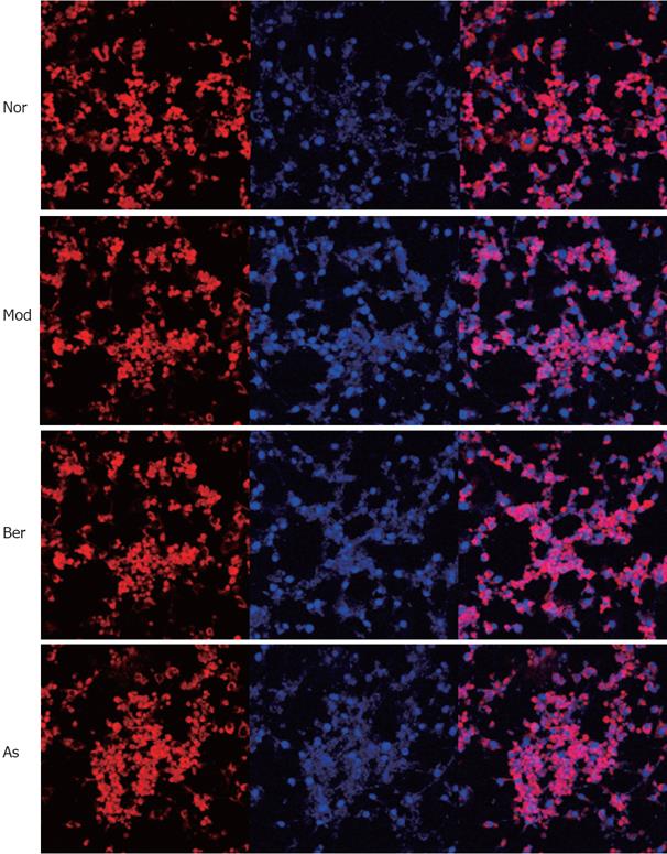
Figure 6 Fully differentiated cells were cultured for 12 h in serum-free DMEM with 0.
2% BSA and then they were, respectively, cultured for 24 h in DMEM containing 1% BSA for 24 h (Nor); or cultured for 24 h in DMEM containing 0.5 mmol/L PA and 1% BSA (Mod); or cultured for 48 h in DMEM containing 10 &mgr;mol/L Ber, 0.5 mmol/L PA, 1% BSA (Berberine); or cultured for 24h in DMEM containing 5 mmol/L aspirin, 0.5 mmol/L PA, 1% BSA (Aspirin). Without FFA stimulation NF-κB p65 is predominately found in the cytoplasm, imparting red color (Nor). While FFA stimulation, NF-κB p65 is translocated into the nucleus, showing pink color (Mod). The addition of 10 &mgr;mol/L Ber or 5 mmol/L aspirin clearly inhibits the FFAs induced NF-κB p65 translocation, as there is hardly any nuclear p65 staining found and in the figure, both red and pink colors can be observed (Berberine and Aspirin).
- Citation: Yi P, Lu FE, Xu LJ, Chen G, Dong H, Wang KF. Berberine reverses free-fatty-acid-induced insulin resistance in 3T3-L1 adipocytes through targeting IKKβ. World J Gastroenterol 2008; 14(6): 876-883
- URL: https://www.wjgnet.com/1007-9327/full/v14/i6/876.htm
- DOI: https://dx.doi.org/10.3748/wjg.14.876


