©2008 The WJG Press and Baishideng.
World J Gastroenterol. Feb 7, 2008; 14(5): 800-802
Published online Feb 7, 2008. doi: 10.3748/wjg.14.800
Published online Feb 7, 2008. doi: 10.3748/wjg.14.800
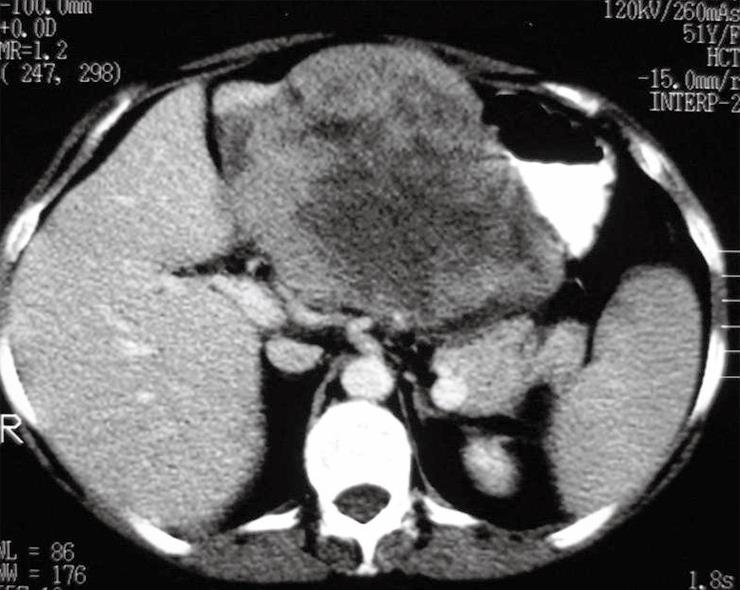
Figure 1 Computed tomography of the abdomen showing a 15-cm heterogeneous, solid expansile, regularly-shaped lesion located in the epigastrium and in contact with the left hepatic lobe and gastric wall.
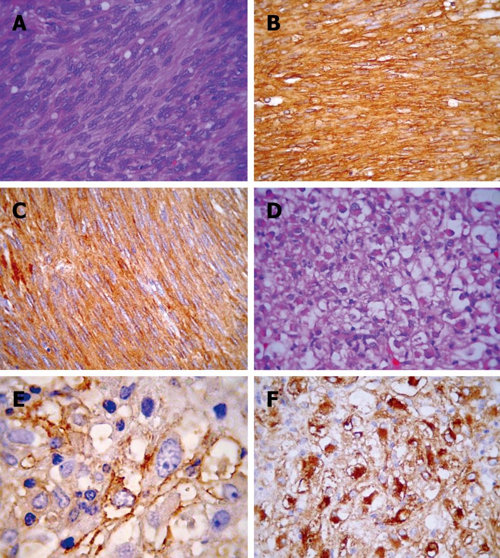
Figure 2 Morphological and immunohistochemical features of gastric GIST (A-C) and hepatic PEComa (D-F).
A: GIST with characteristic spindled bipolar cells with occasional paranuclear cytoplasmic vacuoles; B and C: Strong staining for CD117 and CD34, respectively; D: Hepatic PEComa with a monotonous epithelioid morphology with cytoplasmic clearing that is hardly distinguishable from epithelioid GIST (note scattered cells with rhabdoid cytoplasm); E: A coarsely granular HMB45 reactivity; F: HH35 showing a strong paranuclear cytoplasmic reactivity.
- Citation: Paiva CE, Neto FAM, Agaimy A, Domingues MAC, Rogatto SR. Perivascular epithelioid cell tumor of the liver coexisting with a gastrointestinal stromal tumor. World J Gastroenterol 2008; 14(5): 800-802
- URL: https://www.wjgnet.com/1007-9327/full/v14/i5/800.htm
- DOI: https://dx.doi.org/10.3748/wjg.14.800


