©2008 The WJG Press and Baishideng.
World J Gastroenterol. Dec 14, 2008; 14(46): 7059-7067
Published online Dec 14, 2008. doi: 10.3748/wjg.14.7059
Published online Dec 14, 2008. doi: 10.3748/wjg.14.7059
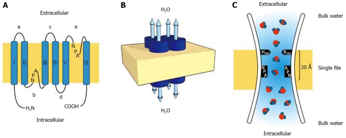
Figure 1 Topology, organization and functioning of the aquaporin water channel molecule.
A: Each AQP monomer consist of six transmembrane domains (I-VI) connected by five loops (a-e) with two NPA boxes shaping the water pore, and the amino and carboxy termini oriented toward the cytoplasm; B: Aquaporins are arranged in tetramers. The water pore does not reside in the center of the molecule, but is formed by connecting loops b and e in each subunit that functions as a unique water pore allowing bidirectional water movement; C: The hourglass model for aquaporin structure. The channel consist of an extracellular and intracellular vestibule containing water in bulk solution joined in the center by a central constriction 20 Å in length where water molecules pass in single file. The a/R constriction delimited by arginine in the position 195 (R195) and histidine in the position 180 (H180) provides fixed positive charges which prevent proton passage. The second constriction is bounded by two asparagine residues from the highly conserved NPA motif. The single water molecule passes through the constriction with no resistance as it forms transient hydrogen bonds with the nearby asparagines.
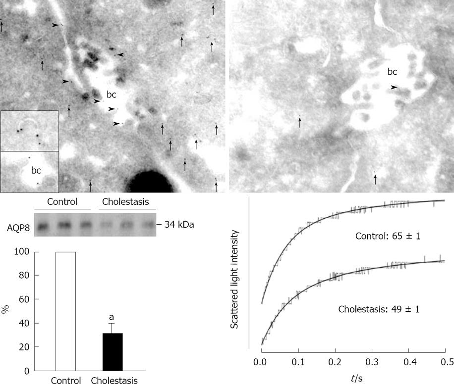
Figure 2 Functional expression of AQP8 in normal and cholestatic liver.
Superior panel: Immunogold electron microscopy for AQP8 in liver from control (left) and 7-d BDL rats (right). Arrowheads indicate AQP8 in bile canalicular (bc) membranes. Arrows indicate AQP8 in the pericanalicular cytoplasm. The upper inset shows an AQP8-containing vesicle, and the lower insert shows AQP8 in the tip of a microvillus and in the intermicrovillar plasma membrane region. (Original magnification X 60 000) Modified and reproduced with permission[57]. Inferior panel: On the left, anti-AQP8 immunoblot of the canalicular plasma membranes from normal and LPS-induced cholestatic liver. On the right, water permeability assessment of canalicular membranes from LPS-induced cholestatic liver. Typical tracings of a time course of scattered light intensity (osmotic water transport), along with single exponential fits in canalicular plasma membrane vesicles in response to a 250 mosM hypertonic sucrose gradient. Calculated Pf values (μm/s) are shown under each curve. Data are mean ± SE from 3 independent vesicle preparations. aP < 0.05 vs conrtol. Modified and reproduced with permission[64].
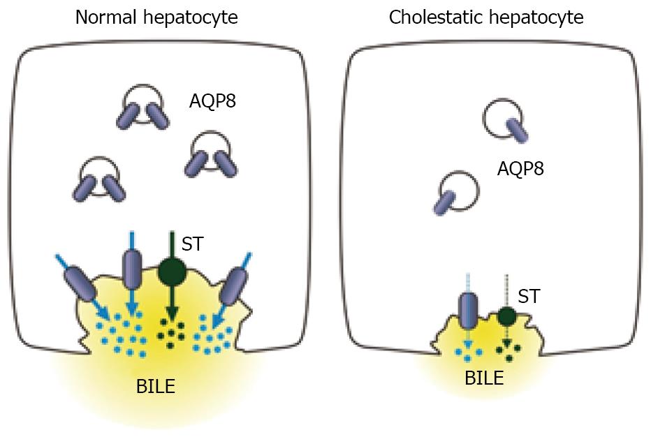
Figure 3 Proposed contribution of hepatocyte AQP8 to the development of cholestasis.
On the left a normal hepatocyte is illustrated with AQP8 expressed at the canalicular membrane domain and in intracellular vesicles. Bile is formed by the active secretion of solute transporters (ST) such as Bsep and Mrp2, which generate the osmotic driving forces for water transport through canalicular AQP8. On the right, a cholestatic hepatocyte is illustrated with decreased expression and functioning of ST. AQP8 is downregulated at the canalicular domain, which impairs the water osmotic permeability thus contributing to decreased bile formation.
- Citation: Lehmann GL, Larocca MC, Soria LR, Marinelli RA. Aquaporins: Their role in cholestatic liver disease. World J Gastroenterol 2008; 14(46): 7059-7067
- URL: https://www.wjgnet.com/1007-9327/full/v14/i46/7059.htm
- DOI: https://dx.doi.org/10.3748/wjg.14.7059


