©2008 The WJG Press and Baishideng.
World J Gastroenterol. Nov 28, 2008; 14(44): 6884-6887
Published online Nov 28, 2008. doi: 10.3748/wjg.14.6884
Published online Nov 28, 2008. doi: 10.3748/wjg.14.6884
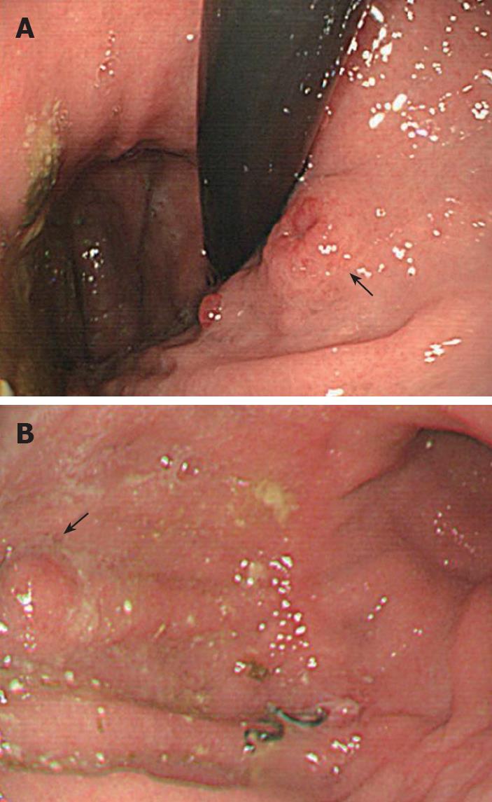
Figure 1 Gastroscopic findings.
A: 1.2 cm ulcerated polyp in the cardia (arrow); B: 0.6 cm polyp on the anterior wall of the corpus near the previous anastomosis (arrow).
Figure 2 Carcinoid tumor.
A: Uniform round-to-oval cells in trabeculae, nests, and gland-like structures (hematoxylin and eosin, × 100); B: Positive immunohistochemical staining for synaptophysin (× 100); C: Negative immunohistochemical staining for CD 34 (× 100); D: Negative immunohistochemical staining for CD 117 (× 100).
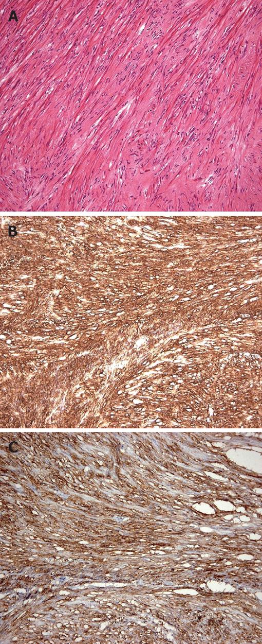
Figure 3 Recurrent gastrointestinal stromal tumor.
A: Composed of spindle cells (hematoxylin and eosin × 100); B: Positive immunohistochemical staining for CD34 (× 100); C: Positive immunohistochemical staining for CD 117 (× 100).
- Citation: Hung CY, Chen MJ, Shih SC, Liu TP, Chan YJ, Wang TE, Chang WH. Gastric carcinoid tumor in a patient with a past history of gastrointestinal stromal tumor of the stomach. World J Gastroenterol 2008; 14(44): 6884-6887
- URL: https://www.wjgnet.com/1007-9327/full/v14/i44/6884.htm
- DOI: https://dx.doi.org/10.3748/wjg.14.6884


