©2008 The WJG Press and Baishideng.
World J Gastroenterol. Oct 21, 2008; 14(39): 6052-6059
Published online Oct 21, 2008. doi: 10.3748/wjg.14.6052
Published online Oct 21, 2008. doi: 10.3748/wjg.14.6052
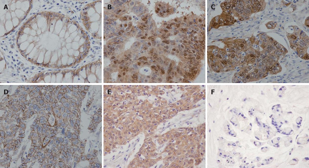
Figure 1 Immunohistochemistry study of beta-catenin in CRC.
A: Membranous staining pattern in normal colonic mucosa; B: Nuclear staining pattern and high OSD in a case with well differentiation histology; C: Nuclear and membranous staining pattern; D Membranous staining pattern; E Cytoplasmic staining pattern; F: Very weak staining intensity in a case of poorly differentiated CRC. (40 × magnification).
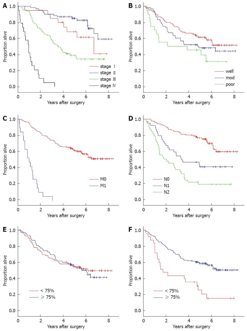
Figure 2 Kaplan-Meier survival curves and log-rank analysis.
A: AJCC staging (P < 0.001); B: Tumor differentiation (P = 0.06); C: Metastatic status (P < 0.001); D: Nodal status (P < 0.001); E: Nuclear staining density (P = 0.59); F: Overall staining density (P < 0.001). Well: Well differentiation; mod: Moderate differentiation; poor: Poorly differentiation.
-
Citation: Wanitsuwan W, Kanngurn S, Boonpipattanapong T, Sangthong R, Sangkhathat S. Overall expression of
beta-catenin outperforms its nuclear accumulation in predicting outcomes of colorectal cancers. World J Gastroenterol 2008; 14(39): 6052-6059 - URL: https://www.wjgnet.com/1007-9327/full/v14/i39/6052.htm
- DOI: https://dx.doi.org/10.3748/wjg.14.6052


