Copyright
©2008 The WJG Press and Baishideng.
World J Gastroenterol. Oct 7, 2008; 14(37): 5760-5762
Published online Oct 7, 2008. doi: 10.3748/wjg.14.5760
Published online Oct 7, 2008. doi: 10.3748/wjg.14.5760
Figure 1 Colonoscopy revealing a semiparent, protruding mucosal lesion covered with normal mucosa with indigested food materials between the lesion and colonic wall (A), which underwent snare polypectomy (B), and inner surface of the lesion filled with seroanguious fluid and yellowish fibroadipose tissue-like materials (C) after the procedure.
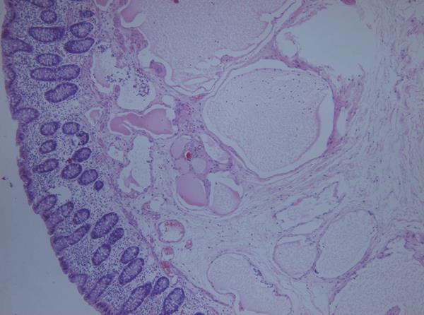
Figure 2 Histopathology showing a cystic lumen covered with a single layer of flat endothelial cells (HE, × 100).
- Citation: Chung WC, Kim HK, Yoo JY, Lee JR, Lee KM, Paik CN, Jang UI, Yang JM. Colonic lymphangiomatosis associated with anemia. World J Gastroenterol 2008; 14(37): 5760-5762
- URL: https://www.wjgnet.com/1007-9327/full/v14/i37/5760.htm
- DOI: https://dx.doi.org/10.3748/wjg.14.5760


