©2008 The WJG Press and Baishideng.
World J Gastroenterol. Sep 21, 2008; 14(35): 5412-5418
Published online Sep 21, 2008. doi: 10.3748/wjg.14.5412
Published online Sep 21, 2008. doi: 10.3748/wjg.14.5412
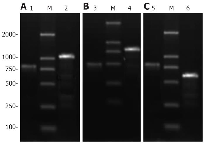
Figure 1 PCR verifying recombinant adenoviruses of Ad-XC (A), Ad-X (B), and Ad-C (C).
M: DL2000; lanes 1, 4 and 5: PAdEasy-1; lane 2: HBV X-HCV C fusion gene; lane 3: X gene; lane 6: C gene.
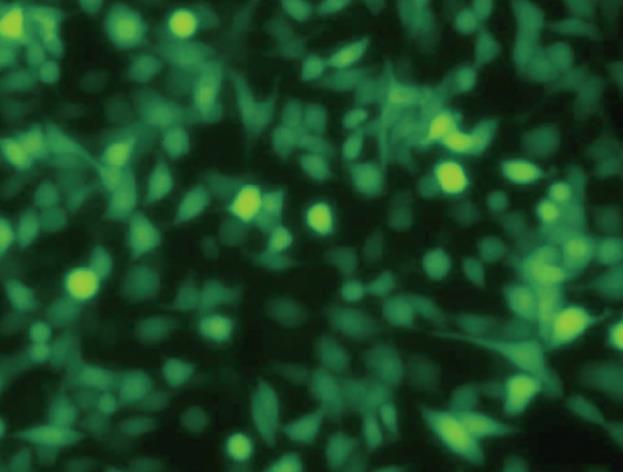
Figure 2 HepG2 cells infected with recombinant adenoviruses (× 200).
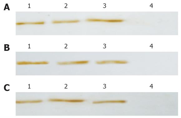
Figure 3 Western blotting displaying expression of fusion protein (A), HBV X protein (B), and HCV C protein (C).
Lanes 1-3: Infected HepG2 cells; lane 4: Uninfected HepG2 cells.
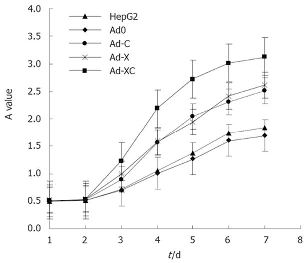
Figure 4 Growth status change in HepG2 cells after infection.
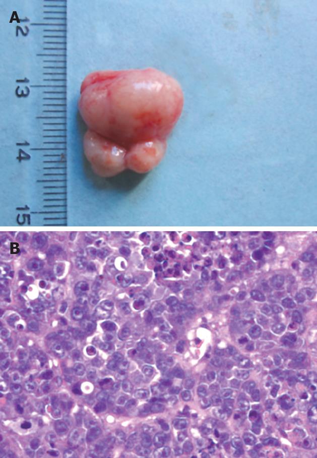
Figure 5 Surgical specimens of tumor tissue (A) and histological examination (B) showing a 1.
5 cm tumor (HE staining, × 400).
Figure 6 RT-PCR revealing mRNA expression of c-myc (A) and relative mRNA expression level of c-myc (B) in HepG2 cells.
M: DL2000; lanes 1-5: HepG2, Ad0, Ad-C, Ad-X and Ad-XC cells (P < 0.05, Ad-XC group vs other groups).
- Citation: Ma Z, Shen QH, Chen GM, Zhang DZ. Biological impact of hepatitis B virus X-hepatitis C virus core fusion gene on human hepatocytes. World J Gastroenterol 2008; 14(35): 5412-5418
- URL: https://www.wjgnet.com/1007-9327/full/v14/i35/5412.htm
- DOI: https://dx.doi.org/10.3748/wjg.14.5412


