Copyright
©2008 The WJG Press and Baishideng.
World J Gastroenterol. Sep 14, 2008; 14(34): 5353-5355
Published online Sep 14, 2008. doi: 10.3748/wjg.14.5353
Published online Sep 14, 2008. doi: 10.3748/wjg.14.5353
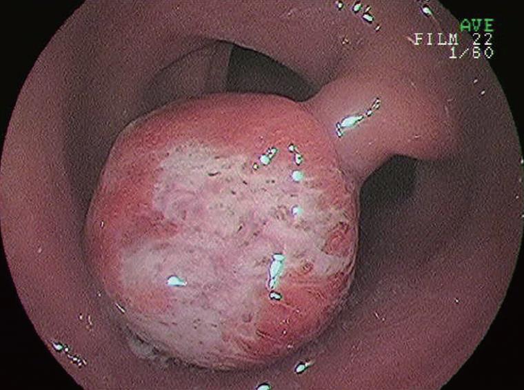
Figure 1 Endoscopy showing a red, hard and spherical peduncular polyp, about 20 mm in diameter, in the descending colon.
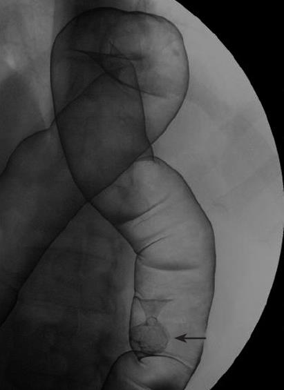
Figure 2 Double contrast radiograph of descending colon showing an about 20 mm peduncular polyp.
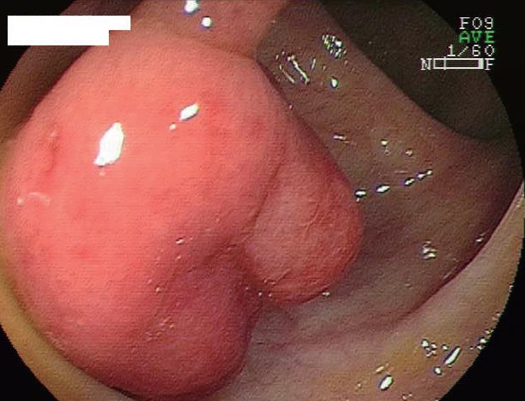
Figure 3 The second colonoscopy showing a healed polyp.
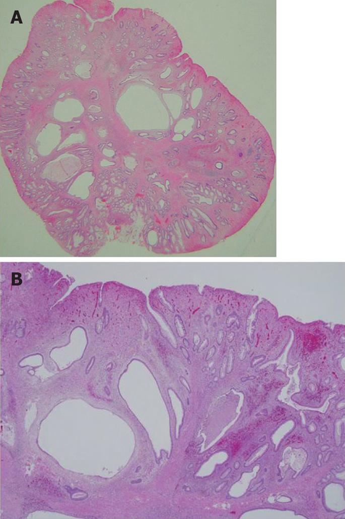
Figure 4 Microscopic findings of the polypectomy specimen.
Low-power view of a cross section showing a stalked polyp containing numerous cystically dilated glands (A) and inflammatory granulation tissue in the lamina propria mucosae and proliferation of smooth muscle (B).
- Citation: Hirasaki S, Okuda M, Kudo K, Suzuki S, Shirakawa A. Inflammatory myoglandular polyp causing hematochezia. World J Gastroenterol 2008; 14(34): 5353-5355
- URL: https://www.wjgnet.com/1007-9327/full/v14/i34/5353.htm
- DOI: https://dx.doi.org/10.3748/wjg.14.5353


