Copyright
©2008 The WJG Press and Baishideng.
World J Gastroenterol. Sep 14, 2008; 14(34): 5331-5335
Published online Sep 14, 2008. doi: 10.3748/wjg.14.5331
Published online Sep 14, 2008. doi: 10.3748/wjg.14.5331
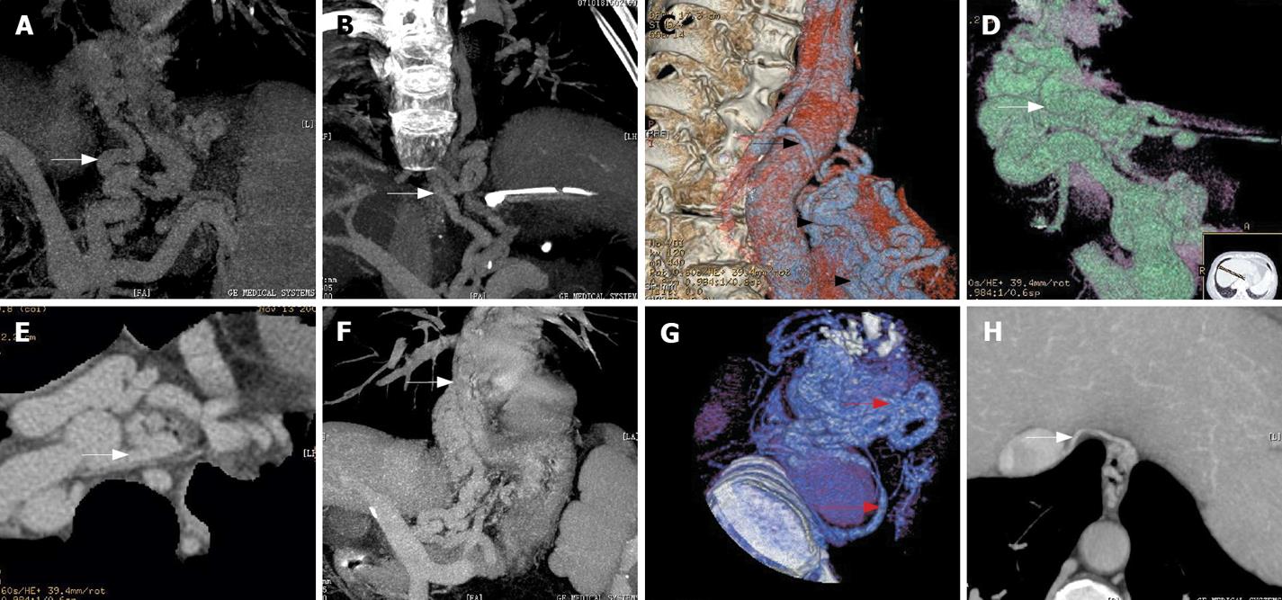
Figure 1 A: Para-EV originated from the posterior branch of the left gastric varices (arrow); B: Circuitous morphological para-EV (arrow); C: Para-EV in reticulatous pattern (short arrow), located at lower section of the esophagus (arrowhead), communicating to hemiazygous vein (long arrow); D: Para-EV communicating to peri-EV (arrow); E: Para-EV communicating to peri-EV directly (arrow); F: Para-EV communicating to peri-EV with peripheral circuitous appearance (arrow); G: Para-EV communicating to peri-EV (short arrow) and the hemiazygous vein (long arrow); H: Para-EV communicating to the subphrenic vein (arrow).
- Citation: Zhao LQ, He W, Chen G. Characteristics of paraesophageal varices: A study with 64-row multidetector computed tomography portal venography. World J Gastroenterol 2008; 14(34): 5331-5335
- URL: https://www.wjgnet.com/1007-9327/full/v14/i34/5331.htm
- DOI: https://dx.doi.org/10.3748/wjg.14.5331


