Copyright
©2008 The WJG Press and Baishideng.
World J Gastroenterol. Jul 14, 2008; 14(26): 4101-4110
Published online Jul 14, 2008. doi: 10.3748/wjg.14.4101
Published online Jul 14, 2008. doi: 10.3748/wjg.14.4101
Figure 1 Summary diagram of established and putative iron absorption pathways in the intestinal enterocyte.
Non-heme iron: All non-heme iron is ultimately taken up from the lumen by DMT1 situated on the microvillus membrane, before joining the labile iron pool in the cytoplasm. Ferric iron must first be reduced to the ferrous form by DcytB before uptake. Ferrous iron in the labile iron pool is then transferred to the circulation by FPN1, which requires hephaestin for oxidation to the ferric form in order to bind to circulating apotransferrin. Heme iron: Heme iron is hypothesized to be taken up by receptor mediated endocytosis. Internalised heme is degraded by HO-2 inside the vesicles, releasing non-heme iron and generating biliverdin. The non-heme iron is then transported to the cytoplasm by DMT1. Heme iron may also be taken up by PCFT/HCP1 directly into the cytoplasm. Intact heme may be transported across the basolateral membrane by FLVCR where it binds circulating hemopexin. Alternatively, heme may be catabolized to non-heme iron and biliverdin by HO-1 located on the endoplasmic reticulum. Any iron released from heme inside the enterocyte, regardless of the mode of uptake, ultimately joins the labile iron pool and is transferred to the bloodstream by FPN1 in the same fashion as non-heme iron.
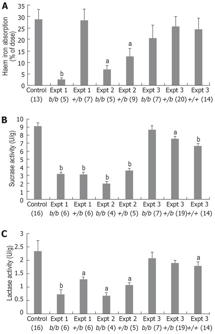
Figure 2 Results from Belgrade rats for heme iron absorption (A), sucrase activity (B) and lactase activity (C) over a series of experiments.
Figures in brackets indicate n values, and data is mean ± SE. Groups marked with ‘a’ or ‘b’ are significantly different from the control group (1-way ANOVA aP < 0.05 and bP < 0.005, respectively). In early experiments 1 and 2, b/b and +/b rats had significantly lower heme iron absorption than Wi controls, initially suggesting a possible role for DMT1. However, sucrase and lactase activity was also lower in b/b and +/b rats indicating a more general defect in the mucosa of the Belgrade strain. In experiment 3, +/+ rats were used as an improved control to account for strain differences between Wi and Belgrade strains, but in this (and subsequent) experiments there was an apparent recovery in both heme iron absorption as well as sucrase and lactase activities. We could find no explanation for this dramatic phenotypic change, making it difficult to correlate DMT1 function with heme iron absorption.
- Citation: West AR, Oates PS. Mechanisms of heme iron absorption: Current questions and controversies. World J Gastroenterol 2008; 14(26): 4101-4110
- URL: https://www.wjgnet.com/1007-9327/full/v14/i26/4101.htm
- DOI: https://dx.doi.org/10.3748/wjg.14.4101


