©2008 The WJG Press and Baishideng.
World J Gastroenterol. Jun 21, 2008; 14(23): 3759-3762
Published online Jun 21, 2008. doi: 10.3748/wjg.14.3759
Published online Jun 21, 2008. doi: 10.3748/wjg.14.3759
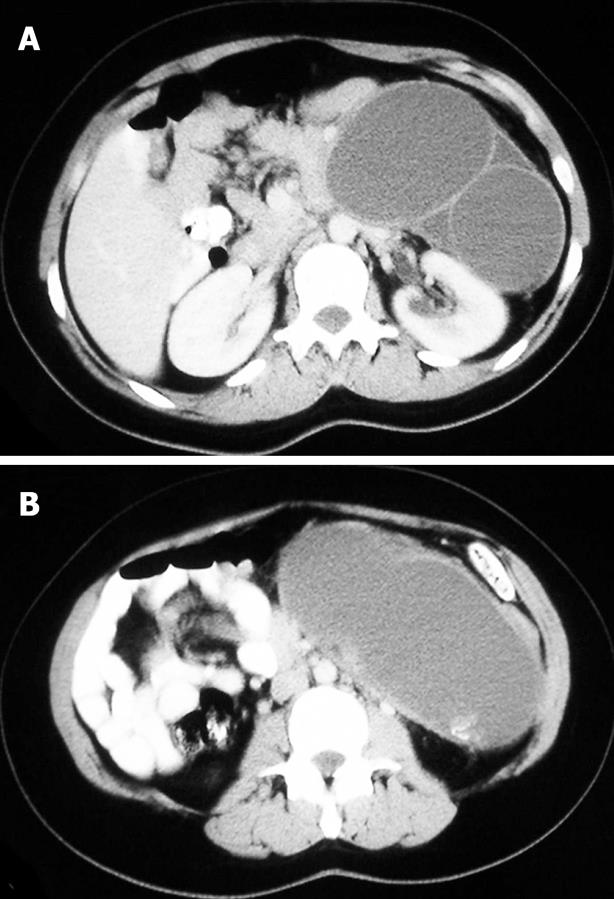
Figure 1 Axial CT scan showing a large bilocular cyst in the region of the body and tail of the pancreas (A) and a second more caudal axial CT slice (B).
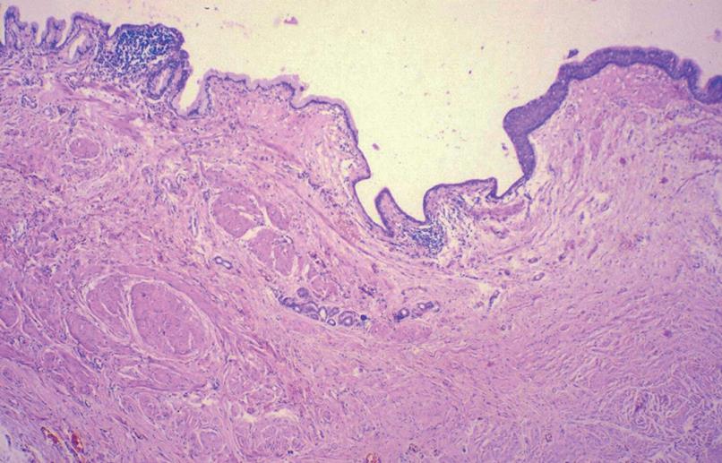
Figure 2 Histological slides showing the full spectrum of epithelia within the cyst lining.
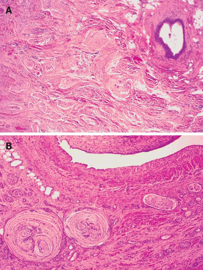
Figure 3 Histological slide showing elements of an abortive muscular layer and focal irregular neuronal hyperplasia in the cyst wall (A) and a more irregular configuration of the cyst with various ganglio-neuronal elements including Paccinian corpuscles (B).
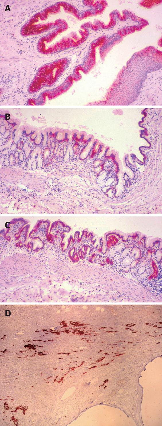
Figure 4 Histological slides showing more specialized epithelia with consistent immunoreactivity to anti-cytokeratin 7 (A), anti-cytokeratin 18 (B), CA19-9 (C), and mixed abundant neuro-ganglionic/neuro-glial elements that were immunohistochemically verified (immunostaining is with the monoclonal antibody to glial fibrillar acidic protein) (D).
- Citation: Colović R, Micev M, Jovanović M, Matić S, Grubor N, Atkinson HDE. Abdominal neurenteric cyst. World J Gastroenterol 2008; 14(23): 3759-3762
- URL: https://www.wjgnet.com/1007-9327/full/v14/i23/3759.htm
- DOI: https://dx.doi.org/10.3748/wjg.14.3759


