©2008 The WJG Press and Baishideng.
World J Gastroenterol. May 21, 2008; 14(19): 3049-3053
Published online May 21, 2008. doi: 10.3748/wjg.14.3049
Published online May 21, 2008. doi: 10.3748/wjg.14.3049
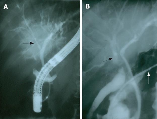
Figure 1 A: Retrograde cholangiogram demonstrating the left hepatic duct and its confluence with the right anterior sectorial duct (arrow, case 7); B: Fistulogram via the drain tube resulted in a retrograde cholangiogram through the transected right posterior sectorial duct (black arrow).
Presence of a nasobiliary tube (white arrow) draining the left hepatic duct (case 7).
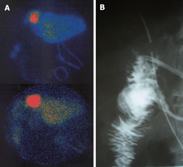
Figure 2 A: HIDA scan demonstrating bile leakage after right extended hepatectomy (case 5); B: Stentogram with intact hepatico-jejunal anastomosis in the same patient (case 5).
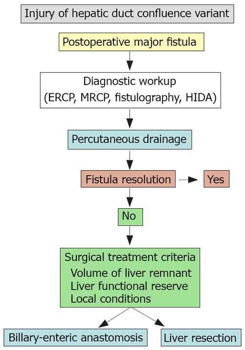
Figure 3 An algorithmic approach for the management of patients with injuries of hepatic duct confluence variants.
- Citation: Fragulidis G, Marinis A, Polydorou A, Konstantinidis C, Anastasopoulos G, Contis J, Voros D, Smyrniotis V. Managing injuries of hepatic duct confluence variants after major hepatobiliary surgery: An algorithmic approach. World J Gastroenterol 2008; 14(19): 3049-3053
- URL: https://www.wjgnet.com/1007-9327/full/v14/i19/3049.htm
- DOI: https://dx.doi.org/10.3748/wjg.14.3049


