©2008 The WJG Press and Baishideng.
World J Gastroenterol. Apr 14, 2008; 14(14): 2280-2283
Published online Apr 14, 2008. doi: 10.3748/wjg.14.2280
Published online Apr 14, 2008. doi: 10.3748/wjg.14.2280
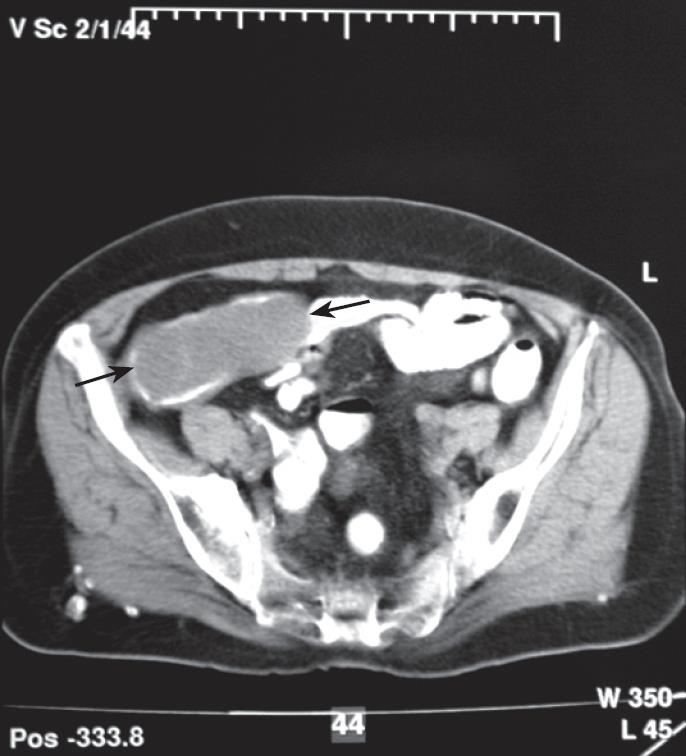
Figure 1 CT imaging of a soft tissue mass indicated by black arrows in the region of the cecum.
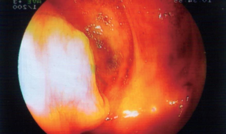
Figure 2 Colonoscopic view of the sub-mucosal mass.
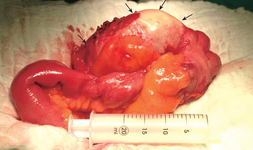
Figure 3 Intra-operative view of the AM.
Arrows indicate the mucine filled appendix.
- Citation: Karakaya K, Barut F, Emre AU, Ucan HB, Cakmak GK, Irkorucu O, Tascilar O, Ustundag Y, Comert M. Appendiceal mucocele: Case reports and review of current literature. World J Gastroenterol 2008; 14(14): 2280-2283
- URL: https://www.wjgnet.com/1007-9327/full/v14/i14/2280.htm
- DOI: https://dx.doi.org/10.3748/wjg.14.2280


