Copyright
©2008 The WJG Press and Baishideng.
World J Gastroenterol. Apr 7, 2008; 14(13): 2055-2060
Published online Apr 7, 2008. doi: 10.3748/wjg.14.2055
Published online Apr 7, 2008. doi: 10.3748/wjg.14.2055
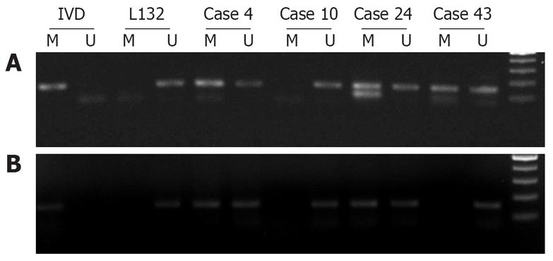
Figure 1 Analysis of p16 promoter hypermethylation in tissue and corresponding serum of patients.
(A) MSP analysis. IVD served as a positive control for hypermethylated DNA and L132 as a positive control for unmethylated DNA. Patients 4, 24 and 43 were hypermethylated, which revealed 150 bp bands with hypermethylated primers. Patient 10 was not methylated. (B) MSP analysis in corresponding sera of samples depicted in A. Patients 4 and 24 were hypermethylated in serum as well. Patients 43 and 10 were not methylated.
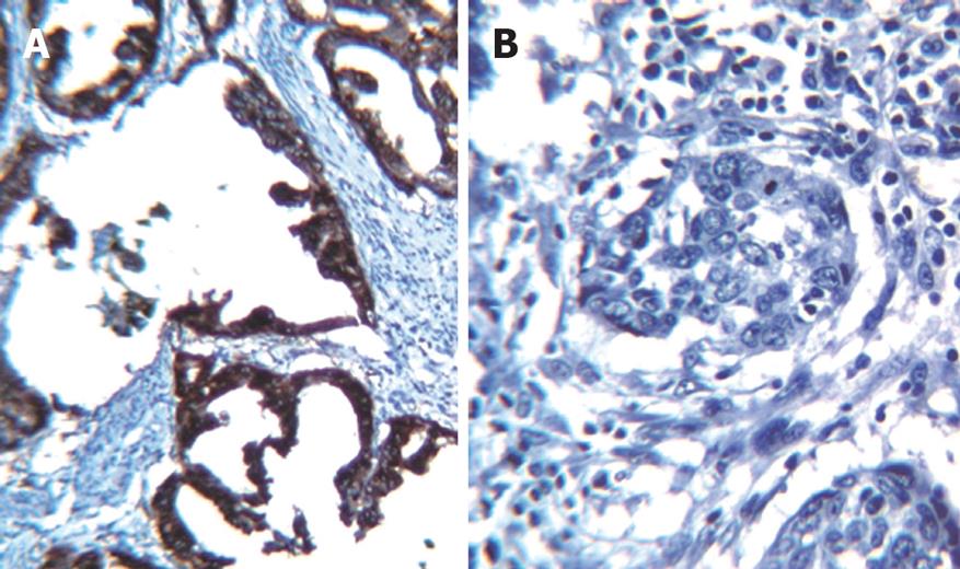
Figure 2 Immunohistochemical staining with monoclonal anti-p16 protein.
(A) Nuclear reactivity showed expression of p16 protein (case 48). (B) p16-negative tumor (case 15) failed to stain due to decreased expression of p16 protein.
-
Citation: Abbaszadegan MR, Moaven O, Sima HR, Ghafarzadegan K, A'rabi A, Forghani MN, Raziee HR, Mashhadinejad A, Jafarzadeh M, Esmaili-Shandiz E, Dadkhah E.
p16 promoter hypermethylation: A useful serum marker for early detection of gastric cancer. World J Gastroenterol 2008; 14(13): 2055-2060 - URL: https://www.wjgnet.com/1007-9327/full/v14/i13/2055.htm
- DOI: https://dx.doi.org/10.3748/wjg.14.2055


