Copyright
©2008 The WJG Press and Baishideng.
World J Gastroenterol. Mar 14, 2008; 14(10): 1504-1509
Published online Mar 14, 2008. doi: 10.3748/wjg.14.1504
Published online Mar 14, 2008. doi: 10.3748/wjg.14.1504
Figure 1 The effect of TL on the growth of SW1990 cells.
SW1990 cells were treated with various concentrations of TL for 6 h, 12 h, 24 h or 48 h and viability was determined by MTT assay.
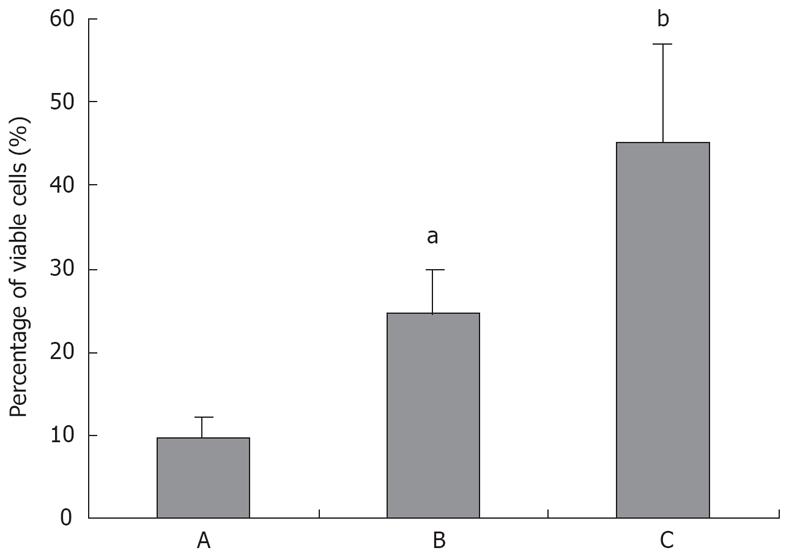
Figure 2 Triptolide induces significant cell death in pancreatic cancer cell line SW1990.
A: TL 0 ng/mL; B: TL 40 ng/mL; C: TL 160 ng/mL. aP < 0.05, bP < 0.001.
Figure 3 TL induced apoptosis in SW1990 cell lines.
SW1990 cells were suspensed in 100 &mgr;L binding buffer and Annexin V/PI double staining was performed, results showed that 40 ng/mL TL indued increased number of apoptotic cells (Annexin V+/PI-).
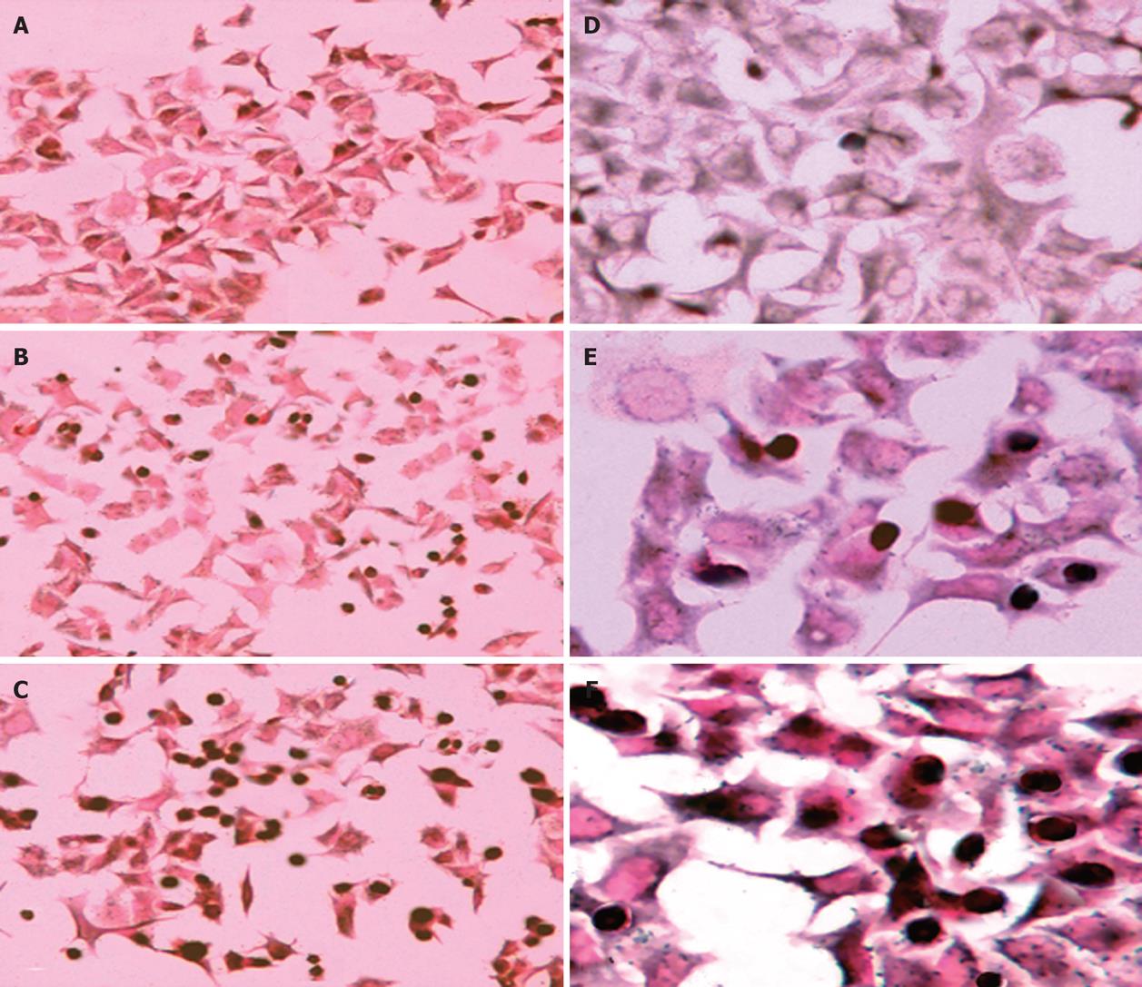
Figure 4 Apoptotic cell death was revealed by TUNEL staining after 24 h (A, B, C magnification, × 200; D, E, F magnification, × 400) of treatment TL.
A, D (TL 0 ng/mL); B, E (TL 40 ng/mL); C, F (TL 160 ng/mL); Apoptotic cells appeared dark after staining.
Figure 5 Detection of Bcl-2, Bax and caspase-3 gene in TL-treated SW1990 cells.
Cells were treated with various concentrations of TL and Bcl-2 ,Bax and caspase-3 genes were analyzed by RT-PCR. A: 1: TL 0 ng/mL; 2: TL 40 ng/mL; 3: TL 80 ng/mL; 4: TL 160 ng/mL; B: 1: Caspase-3; 2: Bax; 3: Bcl-2. bP < 0.01, dP < 0.001.
- Citation: Zhou GX, Ding XL, Huang JF, Zhang H, Wu SB, Cheng JP, Wei Q. Apoptosis of human pancreatic cancer cells induced by Triptolide. World J Gastroenterol 2008; 14(10): 1504-1509
- URL: https://www.wjgnet.com/1007-9327/full/v14/i10/1504.htm
- DOI: https://dx.doi.org/10.3748/wjg.14.1504


