©2007 Baishideng Publishing Group Co.
World J Gastroenterol. Feb 28, 2007; 13(8): 1268-1270
Published online Feb 28, 2007. doi: 10.3748/wjg.v13.i8.1268
Published online Feb 28, 2007. doi: 10.3748/wjg.v13.i8.1268
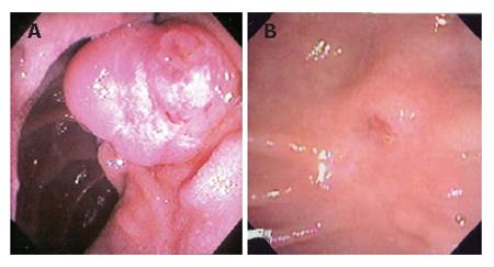
Figure 1 Endoscopic view showing an ampullary tumor (A) and the same area weeks after endoscopic resection of the tumor (B).
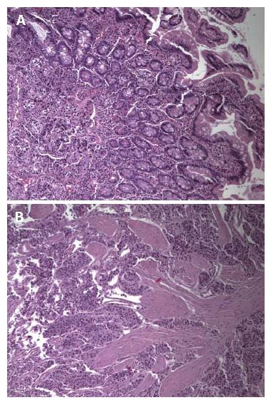
Figure 2 Histological examination of the resected specimen revealing carcinoid in the mucosa and submucosa, composed of discrete solid nests of round tumor cells with central nuclei and occasionally with gland-like lumina (A) and solid nests of tumor cells involving the musclaris propria (B) (HE, x 100).
- Citation: Gilani N, Ramirez FC. Endoscopic resection of an ampullary carcinoid presenting with upper gastrointestinal bleeding: A case report and review of the literature. World J Gastroenterol 2007; 13(8): 1268-1270
- URL: https://www.wjgnet.com/1007-9327/full/v13/i8/1268.htm
- DOI: https://dx.doi.org/10.3748/wjg.v13.i8.1268


