©2007 Baishideng Publishing Group Co.
World J Gastroenterol. Feb 21, 2007; 13(7): 1141-1143
Published online Feb 21, 2007. doi: 10.3748/wjg.v13.i7.1141
Published online Feb 21, 2007. doi: 10.3748/wjg.v13.i7.1141
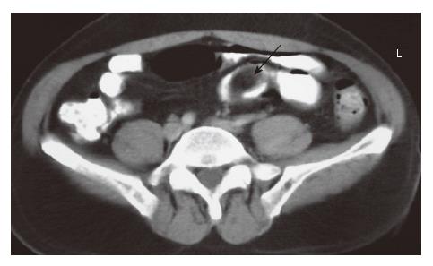
Figure 1 Contrast enhanced CT shows a hypodens, regular contoured, polypoid, 4 cm × 2 cm mass lesion with fatty density.
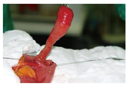
Figure 2 A pedinculated mass lesion within the inverted Meckel’s diverticulum.
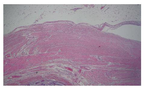
Figure 3 Microscopic appearance of a subserosal lipoma within the inverted Meckel’s diverticulum (HE).
- Citation: Karadeniz Cakmak G, Emre AU, Tascilar O, Bektaş S, Uçan BH, Irkorucu O, Karakaya K, Ustundag Y, Comert M. Lipoma within inverted Meckel’s diverticulum as a cause of recurrent partial intestinal obstruction and hemorrhage: A case report and review of literature. World J Gastroenterol 2007; 13(7): 1141-1143
- URL: https://www.wjgnet.com/1007-9327/full/v13/i7/1141.htm
- DOI: https://dx.doi.org/10.3748/wjg.v13.i7.1141


